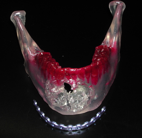


Cysts of the lower jaw can be benign or malignant. But despite the pathology differences they all can cause expansile changes in the bone. When large enough they expand the outer and inner cortical plates of the lower jaw, either pushing out the cortical plates or destroying it in the process.
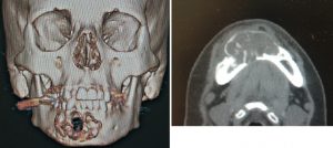 While such jaw cysts are usually diagnosed by conventional x-rays, such as a panorex and 2D CT scans, the planning of the surgery for them should be done by 3D CT scans. Such information provides a better assessment of how large the resection should be, what teeth are involved and their salvageability and how to reconstruct it.
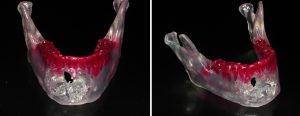 But to improve that visual information and transfer to a tactile one, the use of anatomical models creates a ‘4D’ assessment of the pathology and how to treat it. Anatomical models are made from the patient’s 3D CT scan using stereolithographic layering of acrylic resin.
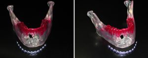 Bring able to see the bone without the limitations of the overlying soft tissues allows the surgeon to determine how to reconstruct it. What is the size of the resultant bone defect, what type and how much bone graft will be needed, and how best to stabilize the remaining bone segments are intraoperative maneuvers that can be determined beforehand. One preoperative maneuver that the anatomical model allows is to prebend a reconstruction plate to the exact curve of the patient’s jaw as well as determine its best placement along the outer cortex of the proximal and distal jaw bony segments.
In today’s 3D Modeling world, plate bending is part of the computer design and such plates are subsequently supplied by the manufacturer ready for surgical implantation.
Dr. Barry Eppley
Indianapolis, Indiana