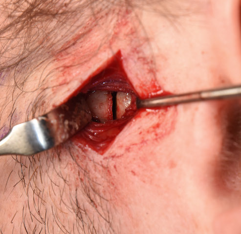


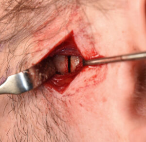
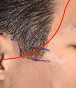
 Classic cheekbone reduction surgery is done by one cuts at the front and back end not the zygomatic bone. While the front bone cut through the zygomatic body is one intraorally, the back bone cut through the end of the zygomatic arch is done externally. This provides the most direct approach to the bone through an overlying skin incision that is typically place at the back edge of the sideburn in men or the preauricular hair tuft in women.
 This bone cut is made with the understanding that the dissection from the skin down to the bone goes right through the territory of the pathway of the frontal branches of the facial nerve. This makes one of the risks of the surgery potential injury to this nerve branch. While this risk is mitigated by blunt dissection after the skin incision is made, it is interesting to know the specific location of the course of these tiny nerve branches.
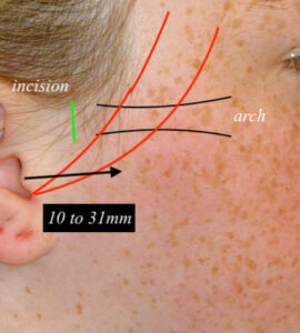 In the August 2020 issue the Aesthetic Surgery Journal an article was published pertinent to this topic entitled ‘Defining a Preauricular Safe Zone: A Cadaveric Study of the Frontotemporal Branch of the Facial Nerve’. Performing a cadaveric study in fresh and embalmed heads, the anatomy of the frontal branches of the facial nerve and its variable branching pattern in the preauricular facial area was studied.The average number of frontotemporal nerve branches crossing the zygomatic arch was two. (2) Beginning from the X point at the apex of the intertragal notch, frontal branches ran over the zygomatic arch at a distance extending from 10 to 31 mm anterior to the tragus, which can be defined as the Danger Zone for frontal branches. Safe Zones A and B are triangular regions located behind and in front of the Danger Zone, respectively.
For the most part the dissection zone for the posterior osteotomy of cheekbone reduction surgery lies just north or at the edge of the most anterior pathway of the frontal nerve branches. This makes the surgical approach safe but not infallible. Thus it is still prudent to use gentle blunt dissection down to the bone with an eye for any tiny nerve branch in the area.
Dr. Barry Eppley
Indianapolis, Indiana