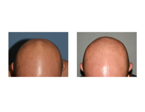
Background: The skull is formed by six separate cranial bones which are initially joined by fibrous tissue known as sutures. The union of these sutures is most commonly recognized by the two large open areas between them that remain after birth, the fontanelles or soft spots. The fontanelles allow for rapid stretching and deformation of the skull bones as the brain expands faster than the surrounding bone can grow together in the first year of life.
The smaller of two most recognized of the baby’s soft spots is the posterior fontanelle. It lies at the junction of the sagittal and lambdoid sutures and is triangular in shape. The posterior fontanelles usually close over by bone growth within a few months after birth. This is done by a process known as intramembranous ossification where the connective tissue of the membrane covering the posterior fontanelle turns into bone.
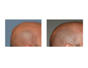
This intramembranous ossification process is not always perfect and can lead to either iincomplete bony fill or actual bony overgrowth. Why this bony growth mechanism may become abnormal is not known. When the bone fill is inadequate a depression occurs and is known as a skull dimple. When the bony fill is excessive an overgrowth occurs and creates a visible hump or bump. Either way the skull contour is not smooth and the bony deformity can be very apparent.
Highlights:
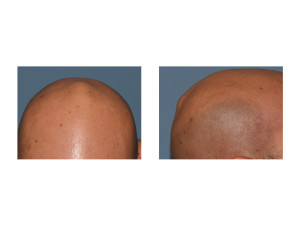
1) One type of abnormally raised skull bump is that which can occur at the vertex or the original posterior fontanelle.
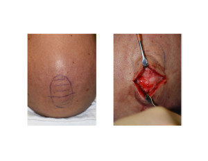
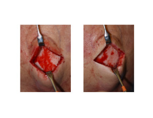
2) The ‘vertex skull bump’ is an abnormally thick segment of bone that permits significant reduction
3) The ‘vertex skull bump’ can be easily reduced through a small scalp incision down to the surrounding skull contour. This is a simple skull reshaping procedure.
Dr. Barry Eppley
Indianapolis, Indiana


