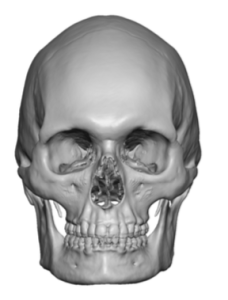
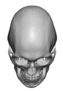
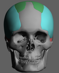
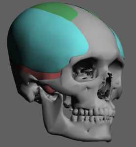
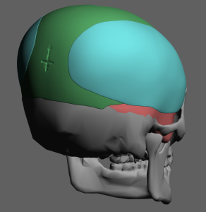
Background: The skull has five basic bony surfaces. The forehead, top and back of the head are exclusively bone surfaces while the paired side of the head is covered by a thick muscular layer. (temporalis muscle) This anatomic difference has great relevance when it comes to performing various skull augmentation procedures. While placing a skull implant on the forehead, top and back of head the placement is always on bone. Conversely augmentation on the side of the head must consider whether implant placement is submuscular (on the bone), subfascial (on top of muscle) or suprafascial. (on top of the deep temporal fascia)
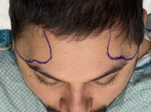
In treating the narrow head shape distinction must be made between an incomplete or complete lack of adequate width. In the incomplete narrow head the main deficiency is central and is located above the ear. It can be treated by placing an implant under the muscle on top of the most convex part of the temporal bone. In the complete narrow head the width deficiency extends into the front and back skull surfaces as well. The forehead is a bit narrow on the sides as well as the same on the back of the head. Augmenting this type of narrow head can not be done by submuscular placement. Rather, because of the fascial attachments along the bony temporal line, an implant augmentation must be placed on top of the deep temporal fascia in order to cross onto the bone on the front and back of the head.
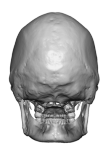
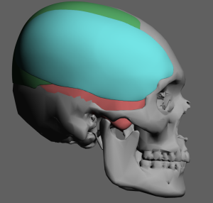
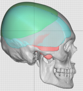
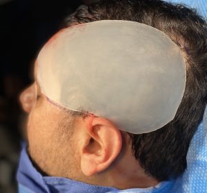
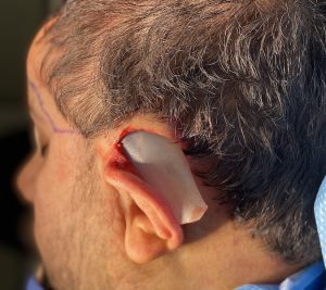
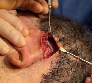
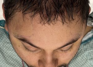
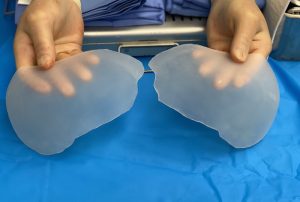
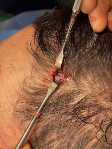
Remarkably such a large head widening implant can be placed through a hidden postauricular and single small scalp incisions. The key to its placement is that it has to be on top of the deep temporal fascia to cross over onto the front and back of the bony skull surfaces. It illustrates that the side of the head is more soft tissue than bone and should be so augmented.
Case Highlights:
1) The narrow head is associated with temporal width deficiencies that often extended into the side of the forehead as well as the side of the back of the head.
2) An extended custom temporal implant can be designed and placed that wraps around the side of the head adding desired width.
3) This extended side of the head implant can be placed through a postauricualr incision behind the ear with one or two smaller positioning scalp incisions.
Dr. Barry Eppley
Indianapolis, Indiana


