

Background: While the nose is composed of bone and cartilage components, its greatest anatomic complexity is in its lower third. The near union of the lower alar cartilages in the midline with the septal support behind it makes for the external size and shape of the nasal tip. The lower alar cartilages is composed of two different legs, a vertical leg (medial crus)which makes up the support of the columella and a higher more obliquely oriented lateral crus that provides support to the nostril. Between the two at the point of maximal tip projection are the domes.
The size and shape of the lower alar cartilages and how they come together any the tip create the projection of the nose. Ideal tip projection has been described by numerous methods including being 55% to 60% of the length of the nose, should be equal to 2/3s of the ideal nasal length and/or the distance from subnasale to the tip should equal to the length of the upper lip.
But diagnosing excessive tip projection is easier that reducing it. Numerous techniques have been described for this nasal tip change, all of which must disrupt the tripod support mechanism. Fundamentally the tip support can be altered at the vertical medial crus, the horizontal lateral crus or right across the tip. (dome truncation) Dome division is generally regarded as the most aggressive of all the tip deprojection techniques and is not a first line approach even if it is the conceptually simplest to do. Generally medial and lateral crus division with overlap is more fundamentally sound.
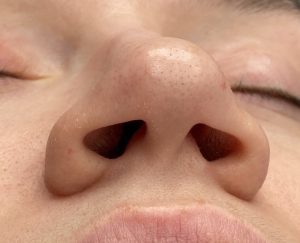
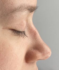
 Case Study: This female had a long nose and desired reduction in its projection, width and increased rotation. There were also some small bumps at the osseocartilaginous junction of the bridge that she wanted reduced.
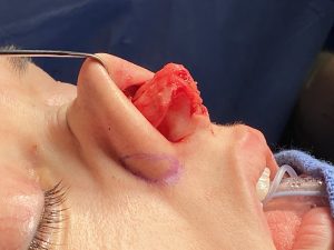
 Through an open rhinoplasty approach the first maneuver was to reduce the bumps on her nasal dorsum which were raised area of the upper lateral cartilage as it joined the nasal bones. With exposure of her long lower alar cartilages, a division at the upper medial crus as well as lateral crus with overlap was done, decreasing , the projection of the nose by 5mms. A cephalic trim was done on each lower alar cartilage with tip narrowing by sutures.
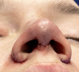 Lastly the wide thick nostrils were reduced by an external wedge reduction technique, placing the scar along the alar-facial groove.
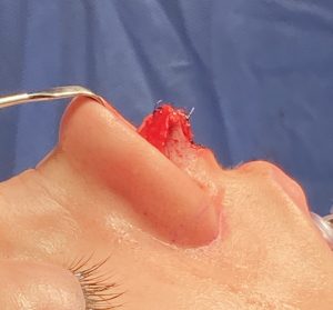
 Her intraoperative result shows the decreased tip projection with increased tip rotation.
Case Highlights:
1) The bulbous long nose is primarily the result of the length, width and separation of the lower alar cartilages.
2) Decreasing tip projection is the most technically challenging dimension of the nose to change in rhinoplasty for which there are several techniques for doing so.
3) With decreased tip projection consideration must be given to the shape of the nostrils which may become disproportionately wide.
Dr. Barry Eppley
Indianapolis, Indiana