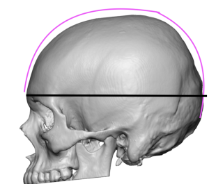
Background: There are a wide variety of aesthetic skull shape deformations that can be divided into either deficiencies or excesses. The excess category may be called protrusions and some are seen in profile where they violate the ‘Golden Arc’ of the skull. This arc is the shape and distance of the head from the nasion (N) anteriorly across the top of the head all the way to the back of the head to the inion. (I) Along the way the arc crosses the bregma (B) and the lambda. (L) These landmarks along this line become linear distances that can be measured and compared by ratios (BI/NB and NI/BI) which ideally are 1.6 or the Golden Ratio.
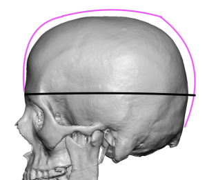
But when the crown of the skull is excessively high it violates a pleasing arc shape. This is due to biparietal skull overgrowth whose reduction depends on how thick the bone is.
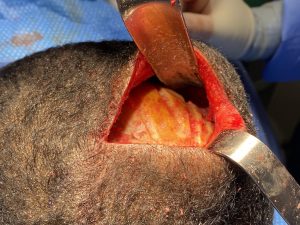
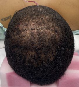
The scalp incision was then closed over a drain. The head dressing and drain were removed the following day.
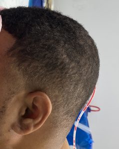
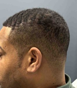
An excessive protrusion of the skull can be visibly reduced provided the bone is thick enough to do so. Most patieents will require a preoperative CT scan to make that assessment.
Key Points:
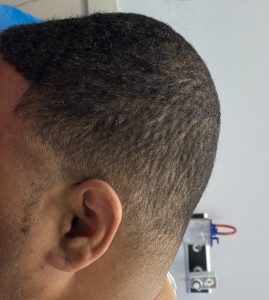
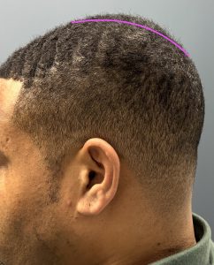
1) The high crown of the skull is due to a thickened parietal skull bone and is clinically seen as an ascending line in profile from front to back with its maximum height posteriorly.
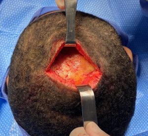
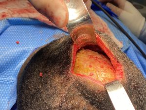
2) A small posterior scalp incision can be used in the prone position to remove the outer cortex of the thickened parietal bone.
3) Parietal or crown of the head reduction restores the normal curvature of the skull in profile and is a procedure which is associated with a rapid recovery.
Dr. Barry Eppley
World-Renowned Plastic Surgeon


