

Brow bone reduction surgery can be performed by two different methods. The most commonly used technique is a bone flap. This is where the anterior table of the frontal sinus is removed, reshaped and then put back. The other technique is bone burring in which the bone thickness is reduced by shaving. Both methods have their place in the properly selected patient which means how much reduction is needed vs the thickness of the brow bone.
For the sake of clarification brow bone reduction can involve two distinct areas of the brow bone. There are inner and outer brow bone prominences and their anatomy is distinctly different. They can be divided by a vertical line that is taken through the pupil of the eyes. What lies inside this line is the inner brow whose prominence is controlled by the development of the underlying frontal sinus. Larger sinus developments create a stronger brow bone prominence which is what occurs in men vs women. Outside this pupillary line is the outer brow bone which is created by the development of the lower frontal bone. Unlike the inner brow bone it is solid bone.
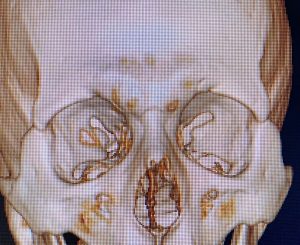
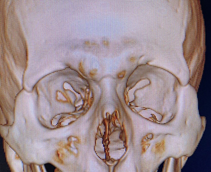
 The relevance of identifying the inner and outer brow bones is that the outer brow bones is always reduced by burring. The often asked and more critical question is whether the inner brow bones can be reduced by burring or needs the setback bone flap method. The answer to this question requires CT scan analysis. In looking at a 3D CT scan it shows the corollary between the bone of the maxillary sinus and that of the frontal sinus. Because the bone is largely filled underneath with an air-filled space, it is quite thin. In the thinnest areas the bone of the medial brow bones, like that of the maxillary sinus, will appear to have ‘holes; in it. In actuality it does not but it is so thin that the reconstructed images can not see it thus leaving it look like it is absent. These holes in the scan represent bone that us about 1mm in thickness.
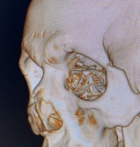
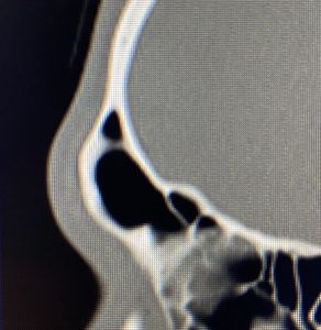 The most prominent part of the brow bone is thicker and it looks so in the 3D CT scan. A 2D CT scan is needed (or even a plain lateral skull film) to measure the thickest part of the medial brow bones. Compared to the edges of the brow bone the most projecting part is usually no more than 3mms in thickness.
Thus for reduction of the medial brow bone prominences if just a few millimeters of bone reduction is needed than s shaving technique can be used. But for more significant reductions the bone flap setback technique is needed.
Dr. Barry Eppley
Indianapolis, Indiana