

Background: Flattening of the back of head can occur on one side or both sides for congenital reasons. When it occurs on just one side of the back of the head it is known as occipital plagiocephaly. It is not, however, just simple limited flattening of one side of the occipital bone. It is well known to be more of an overall twisting of the skull where numerous other craniofacial areas are affected as well. The opposite side of the back of the head, the ipsilateral ear and even the forehead can be altered based on the severity of the deformity.
By far and away the flat side of the back of the head is always the patient’s primary aesthetic skull shape concern. I have used every available onlay cranioplasty material to build up the flat side of the head. The custom skull implant approach using the patient’s 3D CT scan has proven to be superior for a variety of reasons. The exact shape of the deformity correction is determined before surgery, smooth edges of the implant around its perimeter are assured and the implant can be inserted through a small incision due to its flexibility. Because of these benefits it also shortens the time to perform the surgery.
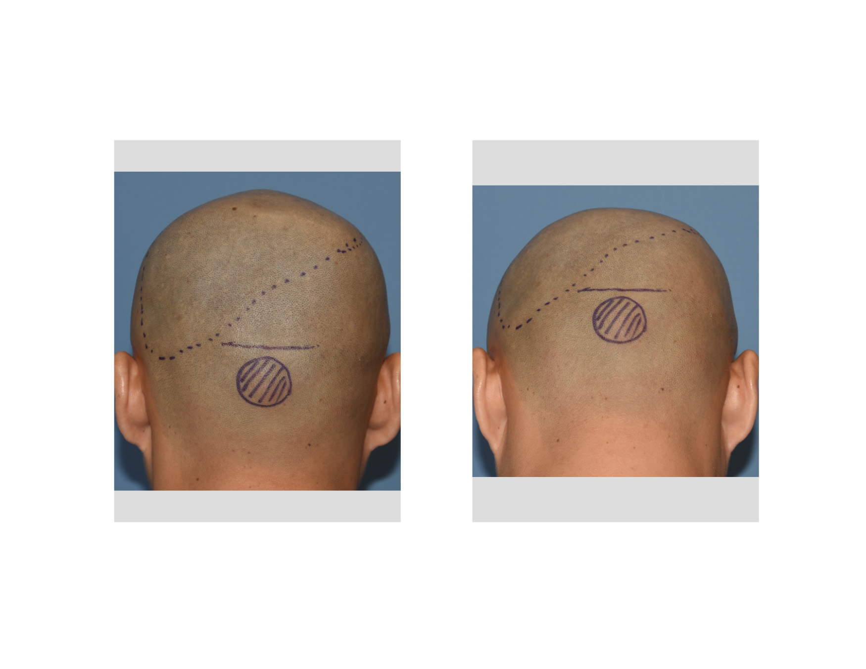 Case Study: This 26 year-old male had long been bothered by the shape of the back of his head. His right side was flat and this was very visible to him since he shaved his head. He also had a moderately sized midline occipital knob which has no known association with occipital plagiocephaly. The area of the implant placement, the occipital knob location and the placement of the horizontal scalp incision was marked on his scalp before surgery.
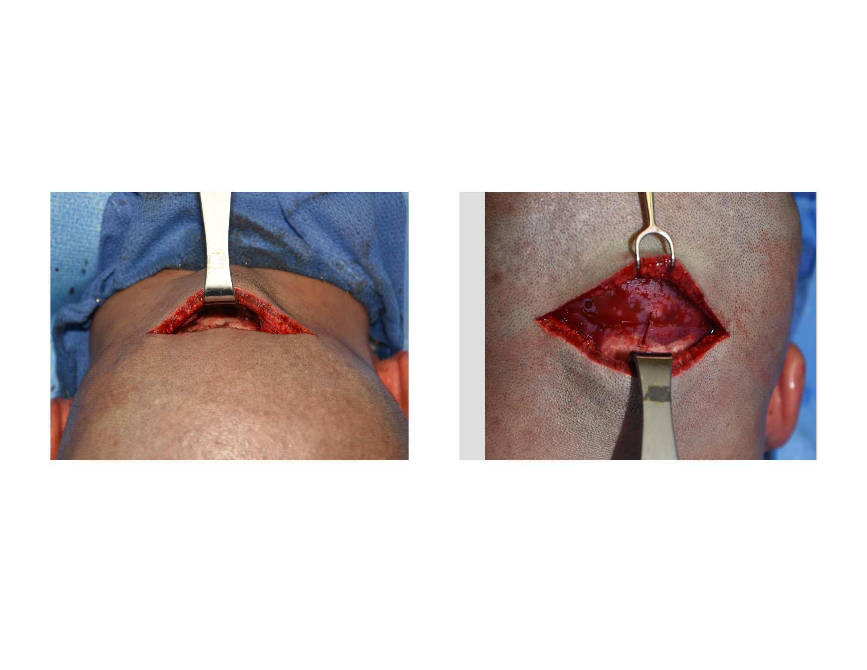
 Under general anesthesia he was placed in the padded prone position which is the only way to perform occipital augmentation when coming from a low incision. Through a 9 cm skin incision, subperiosteal scalp flaps were raised over the location of the implant superiorly and inferiorly down to the occipital knob. The custom occipital implant had multiple perfusion holes made to create through and through tissue ingrowth from the scalp down to the bone into and around the implant after surgery as it healed.
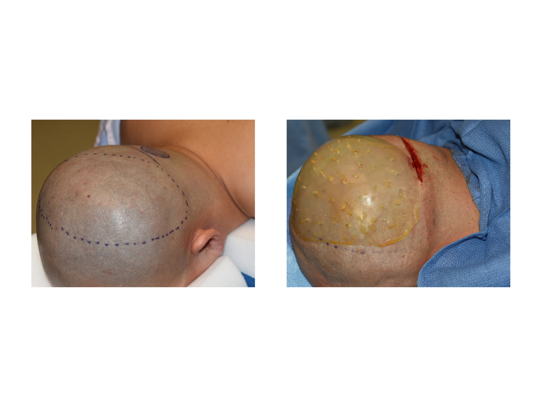
 The custom occipital implant was rolled and inserted through the incision. Once fully inserted and using a preoperative midline mark on the implant, all edges were unfurled to remove all visible edges of the implant and have it lay completely flat. On the bottom side of the incision, the occipital knob was completely burred down to the level of the surrounding occipital bone.
The scalp incision was closed in layers with resorbable sutures. No drain was used. The expected immediate change in the shape of the back of his head was seen. Prior to the placement of a head dressing, greater occipital nerve blocks were done using a long acting local anesthetic. (Marcaine)
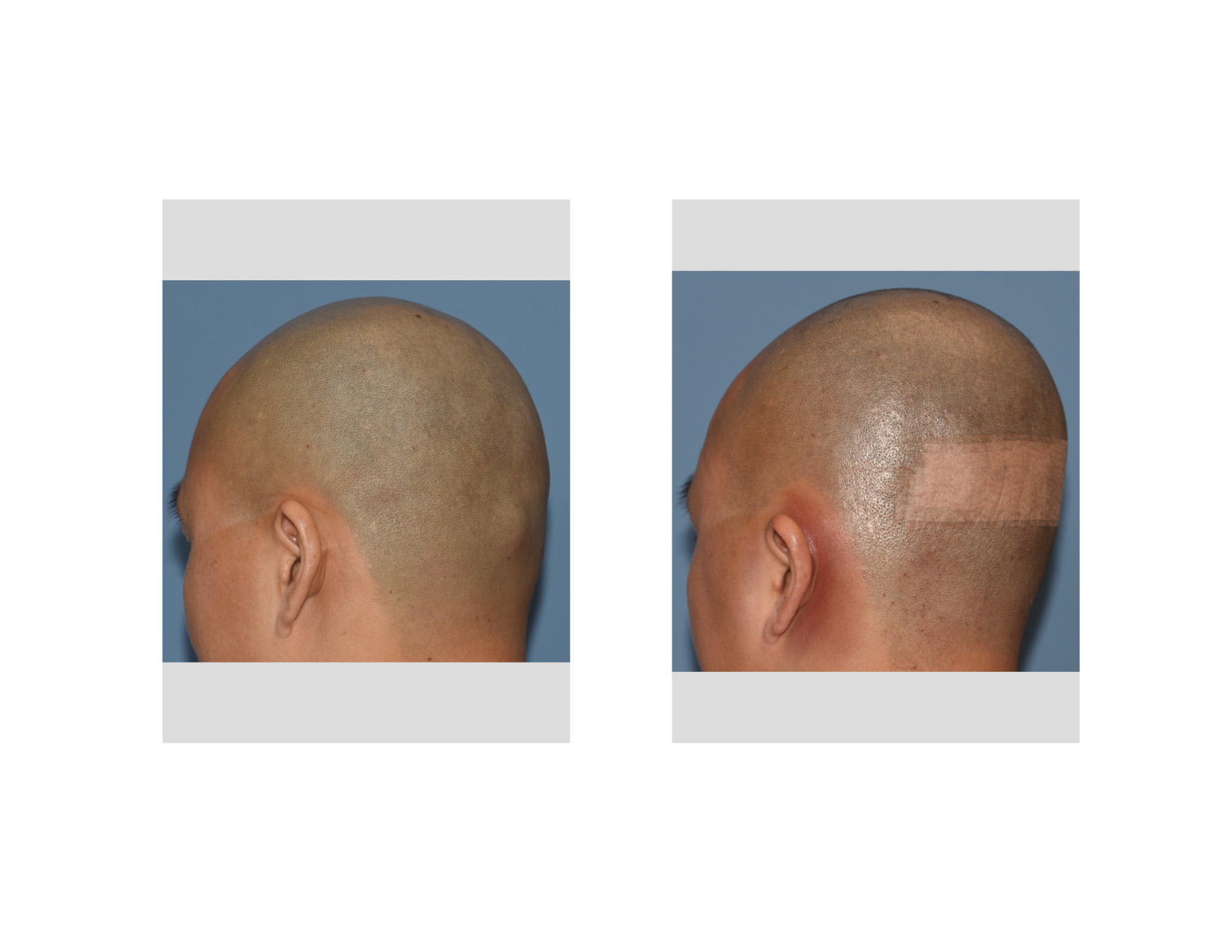
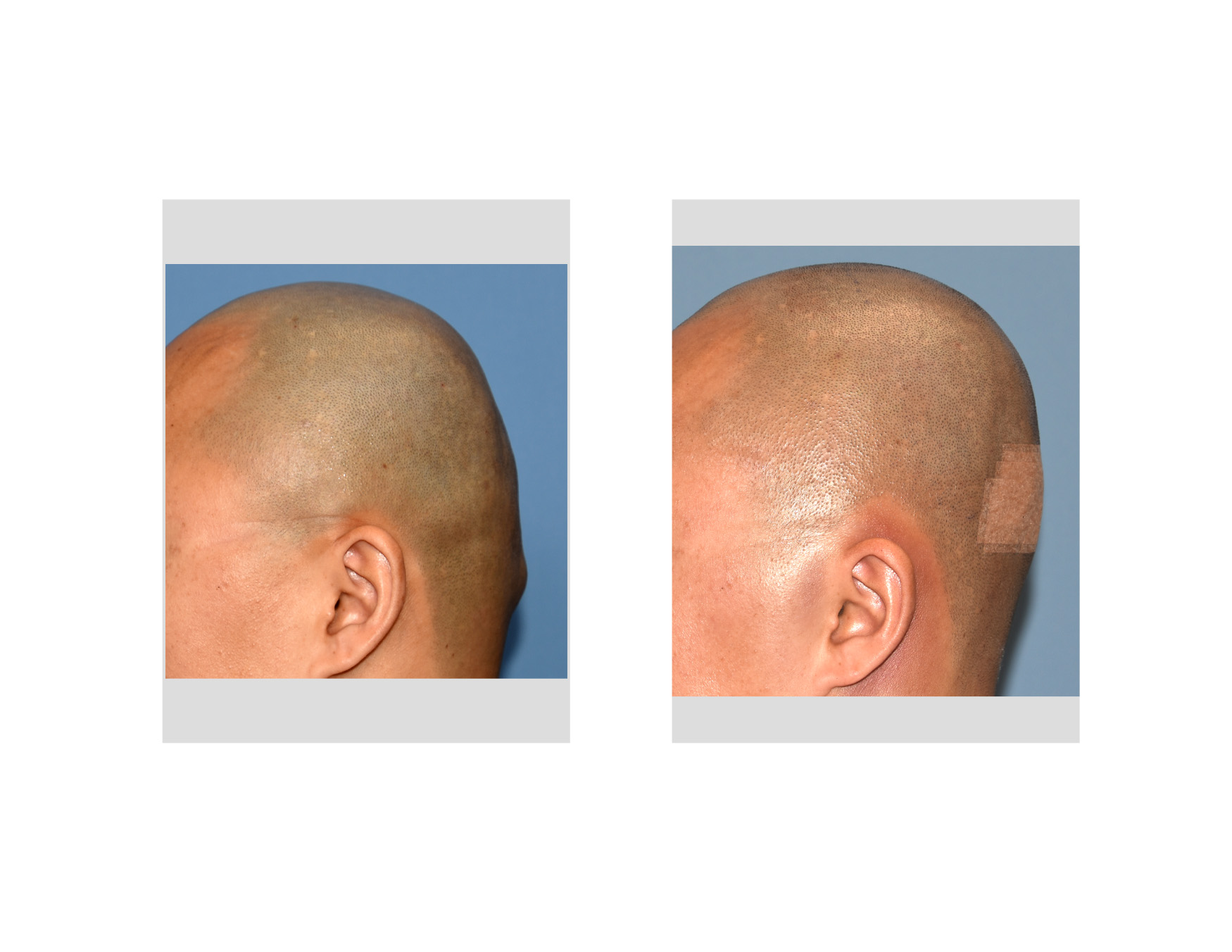
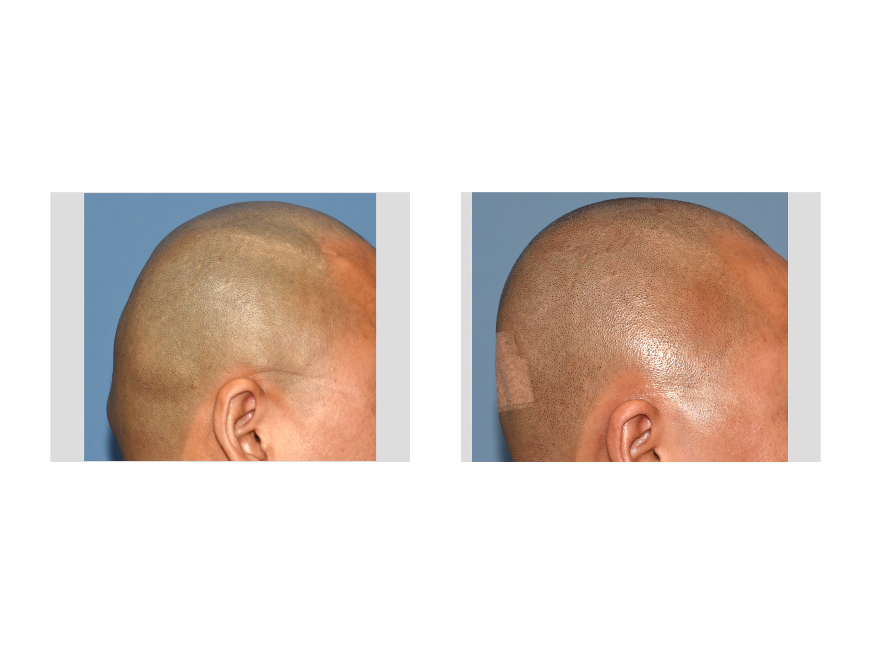 At just one day after surgery, the improvement in his head shape could be seen. Equally importantly, and why I post a one day after surgery picture (only the incision is taped) is that he has no significant bruising and very acceptable swelling. This is partly due to that it is an augmentation procedure where less swelling is always seen and that the tight tissues of the scalp do not allow for large amounts of swelling with limited scalp flap undermining.
Highlights:
1) Congenital occipital plagiocephaly create a visible flattening on one side of the back of the head.
2) One-sided occipital augmentation is most predictably done using a custom occipital skull implant from a 3D CT scan.
3) A custom occipital implant is best introduced through a low horizontal scalp incision on the back of the head.
Dr. Barry Eppley
Indianapolis, Indiana