

Background: The calf or gastrocnemius muscles are a compact and powerful set of lower leg muscles that run from the knee to just above the ankle. It is a bipennate musclke with two known heads. The outer or lateral head starts at the lateral condyle of the femur while the inner head starts from the medial condyle of the femur. The two heads of the muscle join up about midway between the knee and ankle to form a common tendon with the soleus msucle. This common tendon then extends down inferiorly to fix to the heel and is known as the Achilles tendon.
When placing calf implants the anatomy of the aforementioned muscles and tendon is critical. Calf implants are always placed in the subfascial location to sit on top of the muscle. But the subfascial dissection can not extend below the most inferior portion of the muscle where it joins the tendon of the soleus muscle. In the upper half of the lower leg the soleus muscle lies under the gastrocnemius muscles. But as the soleus muscle emerges more superficial at the bottom edge of the gastrocnemius muscles, the overlying fascia becomes very adherent and hard to dissect under.
Most women who seek calf augmentation have a different agenda than that of some men. Women are usually concerned about having a very skinny lower leg that has no definition. (so called ‘chicken legs’) They are looking for some sort of an inner calf ‘bump’ that breaks up an otherwise straight line from the knee to the ankle.
Case Study: This 35 year-old Asian female has long been bothered by the shape of her legs. They were very skinny and has very small calf muscles with a small circumferential calf measurement.
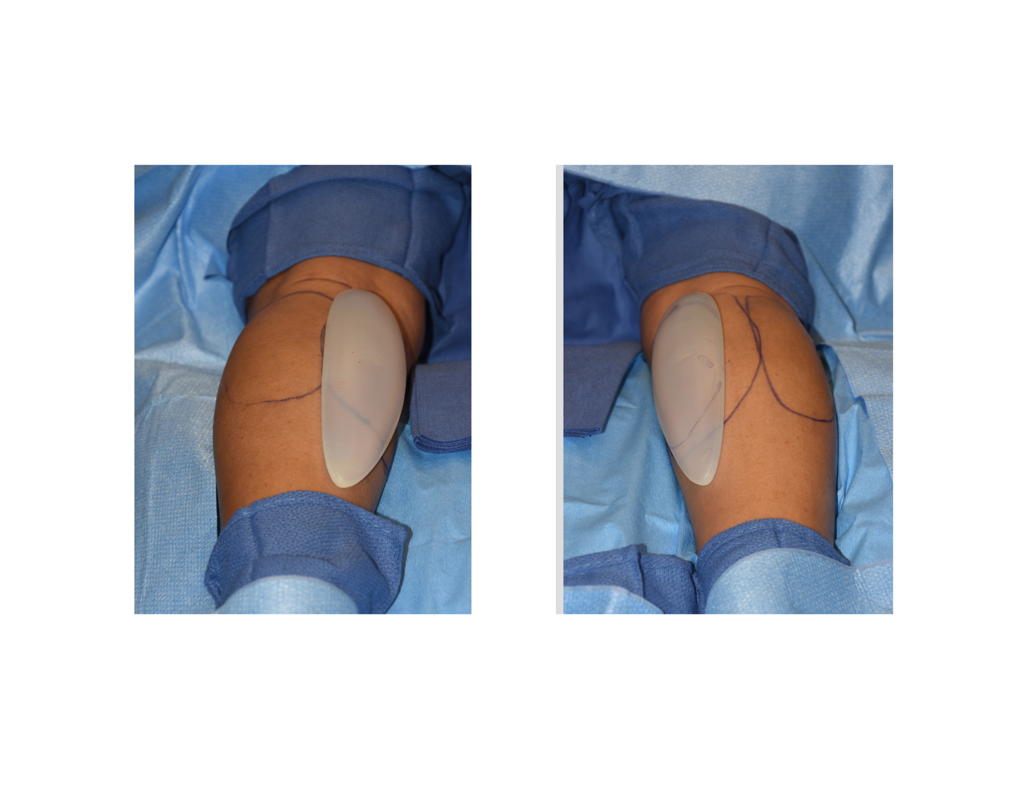
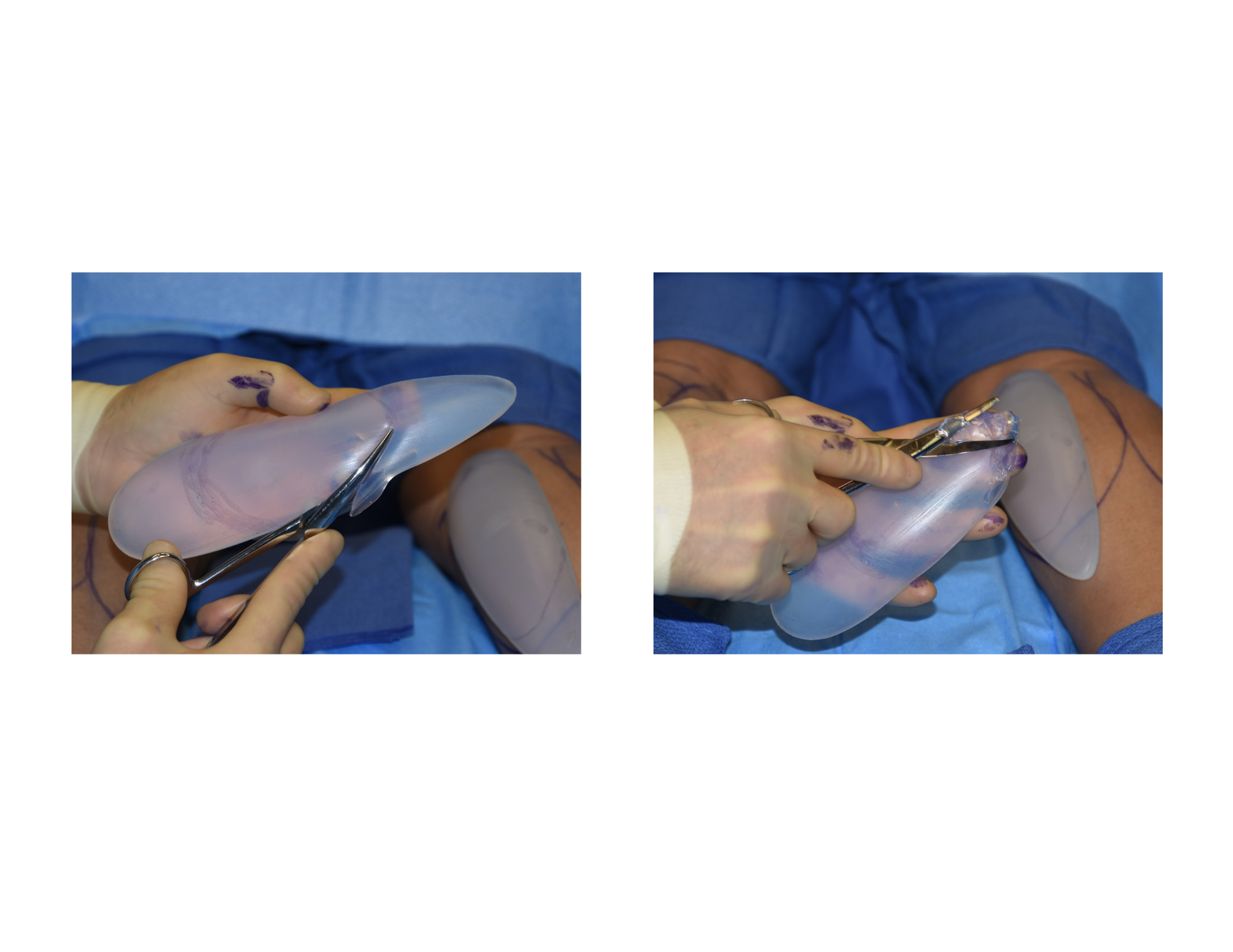 Under general anesthesia in the prone position, a 3.5 cm incision was made in the inner half of the popliteal skin crease. The gastrocnemius fascia was identified, incised and subfascial dissection down with a long broad flat instrument The dissection was done down to the bottom of the calf muscle which was marked beforehand by having her stand on her toes. The standard medium sized calf implants were too long for the length of her gastrocnemius muscles. The calf implants were trimmed to a proper length and then inserted in the subfascial pocket. The fascia was closed over by a fat flap and the skin closed with resorbable sutures in two layers.
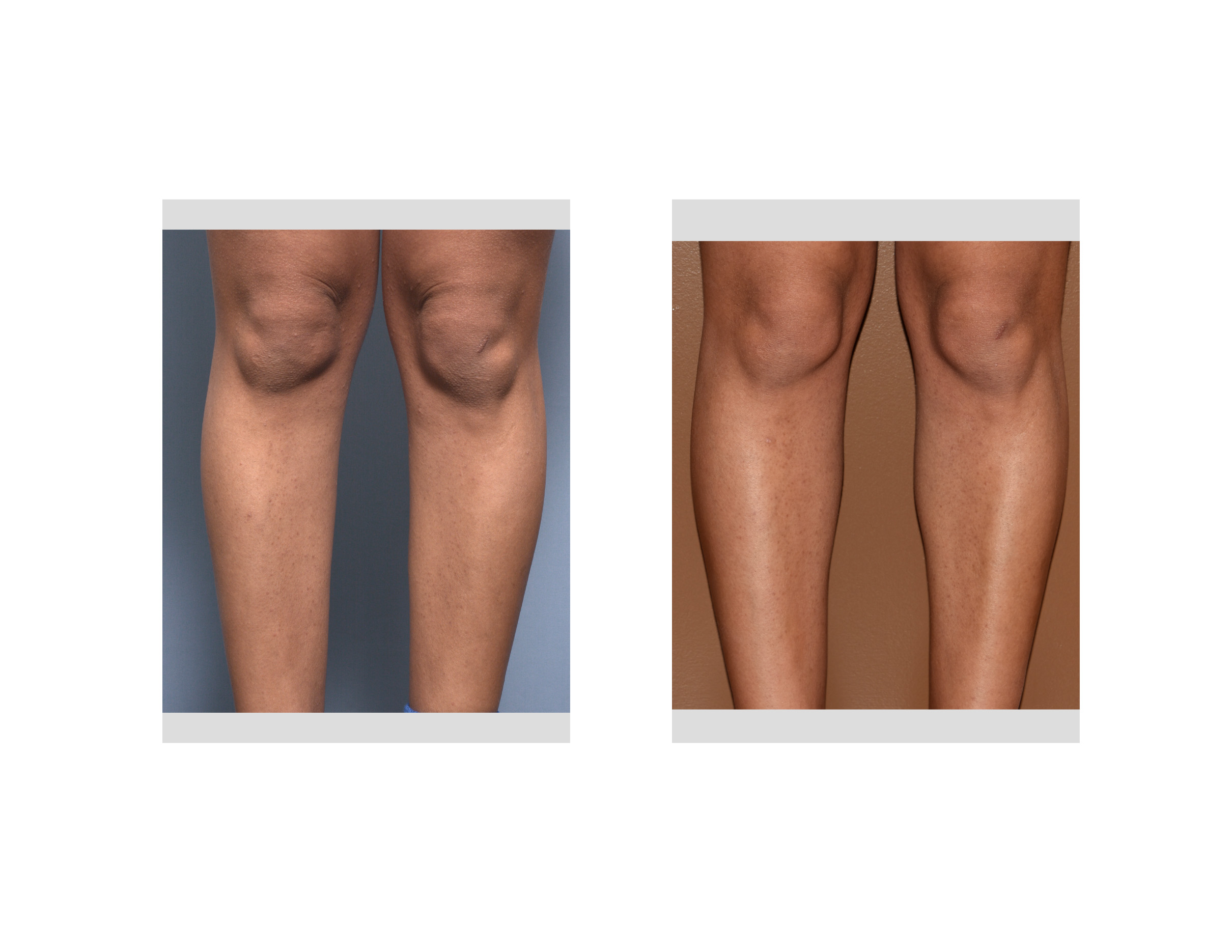
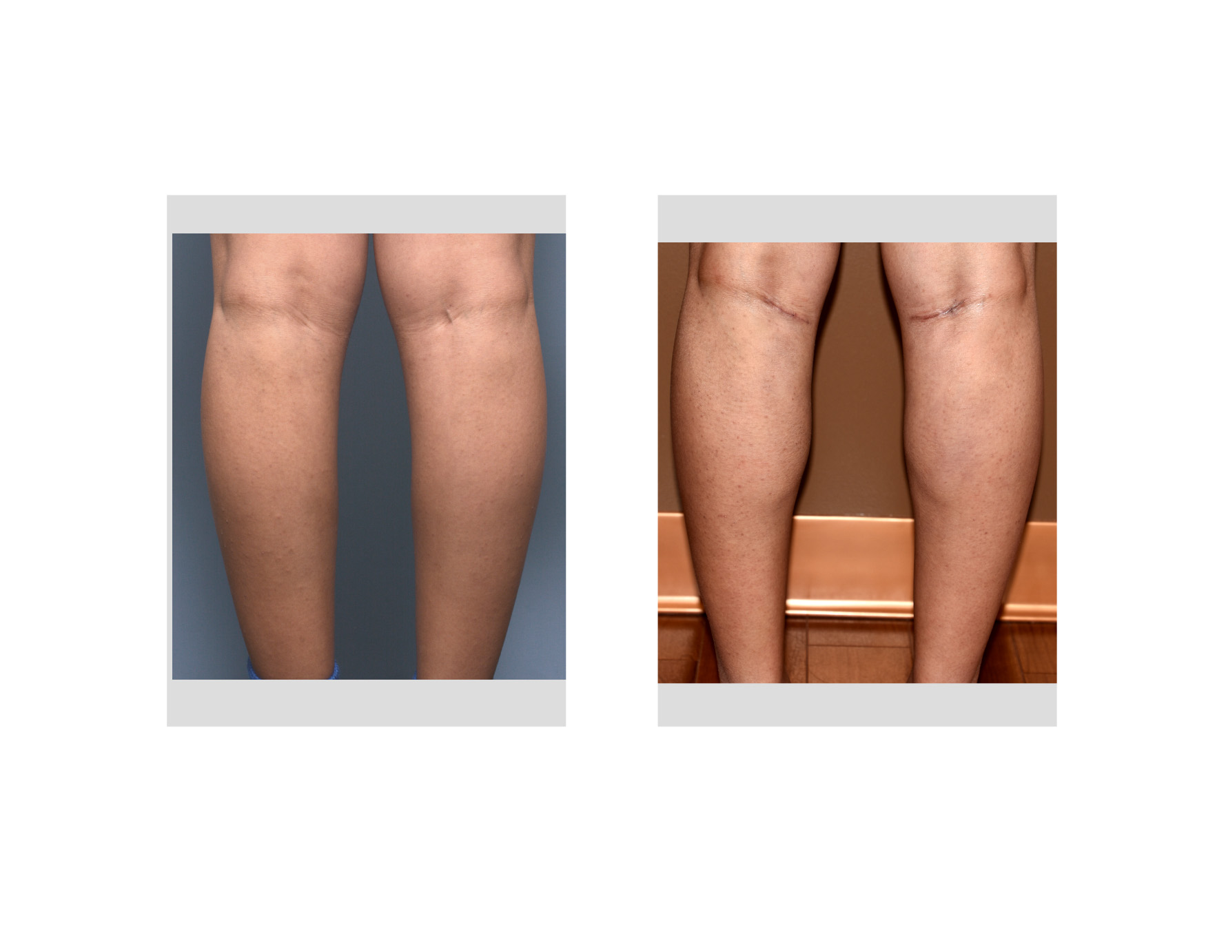 Calf implants in women are designed to create some muscle enhancement in the inner lower leg so it is not just a straight line. In this patient medium calf implants were used whose length needed to be shortened. In hindsight perhaps large calf implants with a wider width would have produced a more significant result. Larger calf implants are longer but shortening the standard length of medium calf implants was needed anyway.
Highlights:
1) Calf implants can be used to augment either in the inner or outer gastrocnemius muscles
2) Most women want to improve the shape of the skinny lower legs through medial or inner calf implants.
3) Inner calf implants are placed in a subfascial plane on top of the muscle through a popliteal crease skin incision.
Dr. Barry Eppley
Indianapolis, Indiana