Background: Loss of the frontal or forehead bone can occur for a variety of reasons, usually from depressed fractures or loss of a craniotomy flap from infection. With removal of the protective bone cover, the brain and its dural covering sit directly up against the skin not only creating an obvious depression but pulsating with each heartbeat. Forehead reconstruction carries the highest aesthetic demands of any skull defect because it is the most visible in a non-hair bearing area and may involve the brow bone and brow ridge area.
There are almost a dozen methods of forehead skull reconstruction from split-thickness cranial bone grafts to computer-generated custom implant pieces. When skillfully done, any of these reconstructive methods will work satisfactorily. Their various advantages and disadvantages change based on the size of the forehead defect. The larger the bone defect becomes the more a synthetic approach becomes an appealing option.
One well established synthetic cranioplasty material for reconstructive use is hydroxyapatite. Consisting of the inorganic mineral content of natural bone, it is highly biocompatible although it does not get replaced by bone. It ends up creating a dense firm bone-like material that blends smoothly into the surrounding bone edges. It does not have the same strength as the normal double cortical layer skull bone but is strong enough to be an adequate skull substitute.
Besides the aesthetics of forehead skull defects, it is the only skull area which is contiguous with the air-filled frontal sinus cavity. This is a potential source of contamination and is a frequent source of forehead infections if a tissue layer is not created between it and the bone reconstruction material.
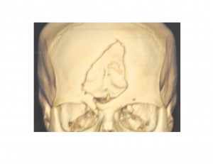
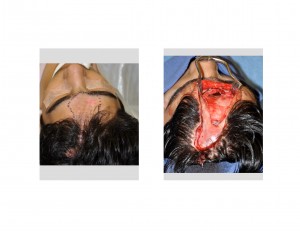
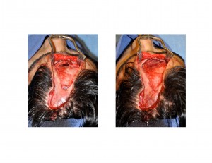
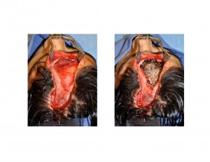
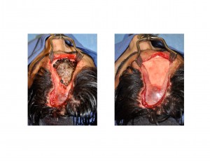
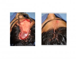
Case Highlights:
1) Reconstruction of the bony forehead can be done by a variety of techniques and hydroxyapatite is a well established cranioplasty material for full-thickness skull defects.
2) Forehead reconstruction which extends down into the brow area must take into account the frontal sinus and have a plan to keep it separate from any implanted material.
3) The properties of hydroxyapatite in a full-thickness skull defect needs reinforcement or a floor to add both strength and a containment method for the material.
Dr. Barry Eppley
Indianapolis, Indiana


