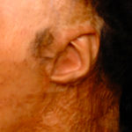
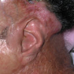
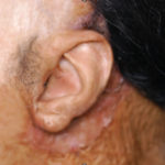
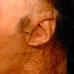
Background: As a projecting facial structure the ear is exposed to a variety of traumatic insults. One such type of traumatic ear injury is that of burns. Most commonly occurring in house and other entrapped fire situations, the sustained exposure to high heat often causes a melted ear appearance. The anterior exposed ear surface often sustains severe and full-thickness burns of the skin and underling cartilage surfaces. The back of the ear is the more protected surface and the postauricular skin and sulcus is often fully preserved or has sustained far less burn injury
While the vast majority of thermal injuries to the ear are due to flames and high heat, another type of burn injury is that caused by chemicals. Chemical burn injuries to the ear are very different from those of high heat. Creating a splash type of contact the burn pattern may not always be on the front of the ear. While the front of the ear is still its most exposed surface, it is possible that the back of the ear can be injured more than the front of the ear. Liquid burn injuries are also somewhat more limited to the contact area without the typical progression of the tissue death associated with high heat damage.
Reconstruction of the burned ear can be challenging because of the loss of normal pliable skin on and around the ear. Often the cartilage structure may be partially or completely useable but scarred and inflexible skin prevents the ear from being separated from the side of the head.


The success of this particular ear reconstruction was largely due to having a preserved cartilage framework. It was the loss of skin surface coverage and the postauricular sulcus that constituted its deformity.
Highlights:
1) Traumatic loss of skin behind the ear causes a fusion of the helical rim to the mastoid skin behind the ear.
2) Tissue expansion and skin grafts can be used to release and reconstruct the postauricular sulcus and nastoid skin surface of the burned ear deformity.
3) The successful reconstruction of a burned ear’s appearance is often based on how well the anterior cartilage structures are preserved.
Dr. Barry Eppley
Indianapolis, Indiana


