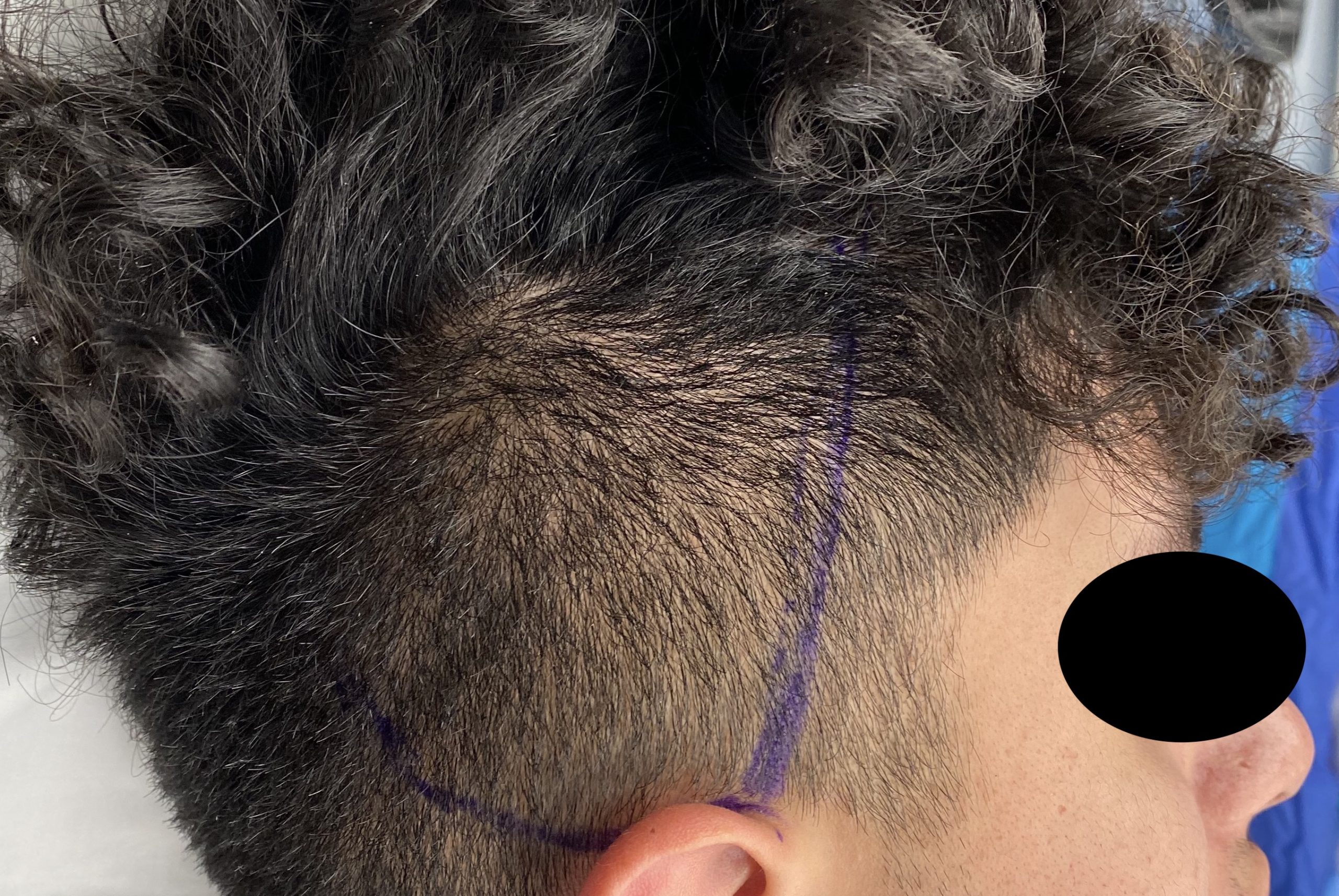
Because of the unique nature of the procedure and lack of any comparative procedure in which to relate, there are numerous misconceptions about temporal reduction. They can be listed and addressed as follows:
HEAD WIDTH REDUCTION REQUIRES BONE REMOVAL TO BE EFFECTIVE
While there is certainly bone on the side of the head, it makes up 50% or less of the total width of the head. The soft tissues make a significant contribution to the side of the head width of which muscle is a major component. But beyond what the ratio of bone to muscle is, there are two specific reasons muscle is the target tissue for reduction. First and foremost it can be removed with no visible scar from an incision behind the ear. Any effort at bone reduction requires a visible incision placed above the ear. Secondly, bone reduction can only take part of the bone (a few millimeters) while muscle removal takes it all. (7 to 10mms thickness) As a result it produces a more effective head width reduction.
TEMPORAL MUSCLE REMOVAL WILL RESULT IN PERMANENT JAW DYSFUNCTION
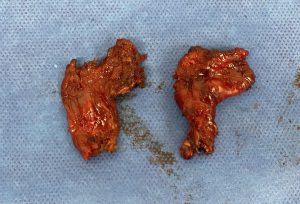
TEMPORAL MUSCLE REMOVAL RESULTS IN PERMANENT NERVE AND BLOOD VESSEL INJURIES
While there are nerves in the temporal region, they all lie in the anterior section. The most important motor nerve, the frontal branch of the facial nerve, actually runs anterior to the temporal hairline between it and the side of the eye. So its preservation is not an issue. The other nerve of way lesser importance, the auriculotemporal sensory nerve, does run through the anterior temporal region above the deep temporal fascia. But that is way anterior to where traditional posterior temporal reduction is done. For the blood vessels there is the posterior branch of the superficial temporal artery that runs over the posterior temporal muscle but this is above the deep temporal fascia while the muscle reduction is done below it.
In conclusion temporal reduction is perfect safely and causes no adverse functional effects despite losing a normal section of muscular anatomy. Thus every patient is evaluated on how likely it will be effective for their head width reduction needs as safety is not an issue.
Dr. Barry Eppley
Indianapolis, Indiana

