

Osteomas are benign bony tumors of the skull that are relatively common, occurring in up to 10% of the population. The most common osteoma is the peripheral type which emanates from the outer surface of the skull. They develop through centrifugal growth from the periosteum with trauma and vessel bleeding being the most recognized cause. (particularly when it occurs at a young age) But most forehead osteomas in my experience have no recognized traumatic origin.
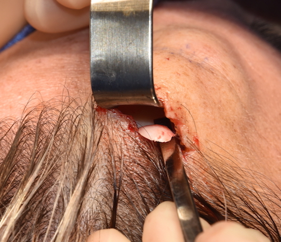
The removal of forehead osteomas should almost always be done without using a direct overlying incision. While a direct incision is the easiest surgical approach it leaves a scar which could potentially be as aesthetically distracting as the osteoma is. They direct incision in the the older patient who has existing deep horizontal wrinkles or in the bald male may be an acceptable approach. But in almost all other cases leaving the exposed forehead free of any scar is the preferred method.
In the November/December 2020 issue of the Journal of Craniofacial Surgery an an article on this topic was published entitled ‘Endoscopic Forehead Approach for Minimally Invasive Frontal Osteoma Excision’. In this paper the authors describe a patient case who underwent endoscopic removal of a forehead osteoma using two 1 cm incisions behind the hairline. No complications from doing so occurred. The endoscopic technique offered good visibility and permitted safe dissection with minimal bleeding. The outcome achieved with endoscopic technique could be the first-line surgical treatment of benign bone tumors of the frontal area, offering more advantages and better results than the conventional surgical approaches.
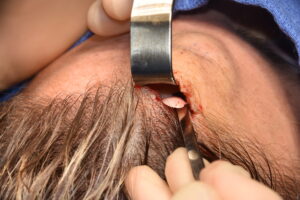 In 2020 a case report using endoscopic removal of a forehead osteoma as a sentinal technique seems dated. This is what I would consider the standard approach and has been so for some time. Rather than a two incision technique it can be done through a single incisional port in my experience in most cases. If the osteoma is in the upper third of the forehead a small hairline or slightly retro hairline incision can be used with direct visualization rather than though endoscopic guidance.
Forehead osteomas should not be confused with forehead horns…which they frequently are. Forehead horns are overdevelopment of the frontal eminences and often occur bilaterally in the upper forehead. Because of their broader base and lack of an edge they can not be removed by an endoscopic technique. They require a burring reduction done through an incision at or behind the hairline directly above them.
Dr. Barry Eppley
Indianapolis, Indiana