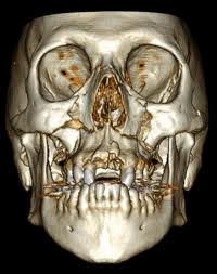
The anatomic structure of the face is well known to change with aging from the outer skin down to the bone. This recognition of how the face ages provides for more effective treatment strategies. While once skin pulling and tightening was the only treatment approach, the understanding of the loss of fat with aging has led to the addition of fat grafting as part of anti-aging facial surgery.

While the bony changes with aging has been studied before, this study is relevant because it assessed the same patients over different time periods. Opening up of the glabellar and maxillary angles as well and increases in pyriform height and width are signs of bone loss/atrophy. Such midface changes can be more severely affected with the loss of teeth although this was not specifically evaluated in this study. (I assume their patients had a reasonably intact maxillary dentition)
The real relevance of this study is whether some patients may benefit by bony augmentation as part of their anti-aging facial surgery. Specifically midface augmentation of the premaxillary-paranasal region may be considered. This could be especially helpful for the patient with deep nasolabial folds and a recessed nasal base.
Dr. Barry Eppley
Indianapolis, Indiana


