

Augmentation of the calfs is most effectively done using implants. The placement of calf implants is on top of the gastrocnemius muscles underneath its fascial covering. Placed through small incisions in a skin crease behind the knee, the fascial plane is relatively easy to find and enter. There are few anatomic structures of which to be aware at this superficial location on the back of the lower leg.
The one anatomic structure to be cognizant of during this direction is the sural nerve. This is a sensory nerve that runs down the middle of the calf to the ankle where it turns around the lateral malleolus to the side of the foot. It supplies sensation to the skin of the side of the foot and ankle. While commonly perceived to run down primarily down between the inner and outer gastrocnemius muscles, a closer look at its anatomy reveals that it is not quite so simple.
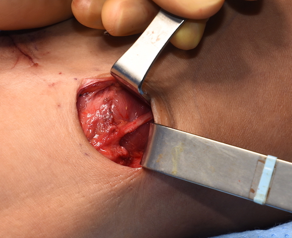
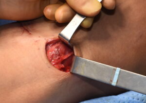 The sural nerve is the fusion of the medial sural cutaneous nerve (MSCN, from the tibial nerve) and the lateral sural cutaneous nerve. (LSCN, from the common fibular nerve) How these two nerve branches come together make for a lot of variability in the pattern of the sural nerves as they cross over the calf muscles. Most commonly the MSCN crosses over the outer calf muscle. This is what is seen in this intraoperative picture of the dissection for the placement of a calf implant to the outer or lateral head of the gastrocnemius muscle. Right in the middle of the dissection runs the MSCN. The LSCN can also run down over the posterolateral surface of the outer calf.
In numerous anatomic dissections that I have done the main branch of the sural nerve is seen to run down the middle between the two heads of the calf muscles. But it is important to recognize that this does not always occur. Branches of the nerve are well known to occur in and around the outer calf muscle as well.
Since the sural nerve is a pure sensory nerve loss of its function only results in some numbness to the side of the foot. But with the introduction of a subfascial calf implant in the proximity of a sural nerve branch, a compression neuropathy can potential occur. (chronic pain) Whether this came from injury to the nerve during dissection or from compression by the implant is never completely known. But removal of the implant is the only treatment for it.
Dr. Barry Eppley
Indianapolis, Indiana