

Background: The ear due to its angulated position from the side of the head is exposed to multiple types of injuries. Most of the these ear injuries are either lacerations (common) or avulsive (far less common) in nature. In either case there are ample normal and supple tissues on or around the ear to perform its reconstruction.
The far less common and most challenging of all ears injuries to repair is that of the burned ear. The significantly burned ear presents a classic pattern of injury with loss of the outer helical framework (looks like it was melted) as well as burned skin in and around the remaining ear. Once primary burn care is completed much of what surrounds the residual ear may be skin grafted. The challenge becomes trying to rebuild an ear which is both stiff, scarred and lacks any normal surrounding tissues.
The main components of any ear reconstruction are cartilage for the framework and a vascularized skin/soft tissue cover of it. Rib is always the source of the cartilage and is available in abundance. It is the vascularized cover of the rib cartilage reconstruction of the ear framework that is the challenge.
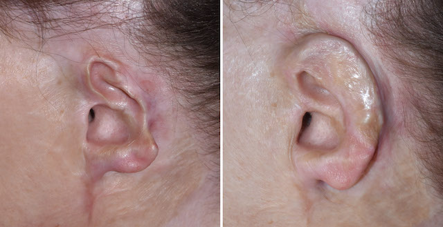
 Case Study: This female suffered a severe 50% total body surface area burn injury that also involved the face, ears, and scalp. After she has been through all of her primary burn care and other reconstructions (several years later) she presented for left ear reconstruction. (her right ear was uninjured) Her goal was not only to rebuild the ear but also to provide a rest for her eyeglasses.
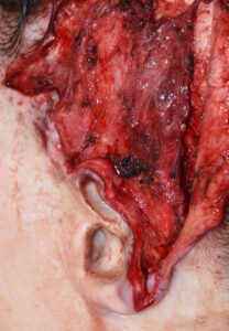 Under general anesthesia a first stage ear reconstruction was performed by initialing removing the scarred skin graft and developing a viable temporal fascial flap.
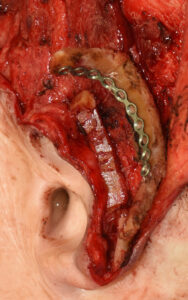
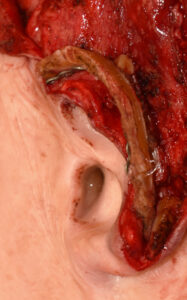 The the harvest of a rib graft from the left subcostal region (rib #8) was done. The cartilage was very hard and partially calcified. (perhaps due to her age and the burn injury) It could not easily be bent to make for a curved helical rim framework. To take a brittle piece of rib cartilage and bend it into a smooth unfractured helical rim shape a small metal plate was placed on its inner surface and the rib sutured to it throughout its length to create the bend.
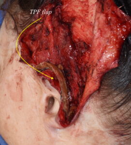 The rib graft was covered by raising the temporoparietal fascia underneath the prior skin grafted scalp. Fortunately the superficial temporal artery flap was intact. The flap was turn down over the ribcartilage and its inner surface (not the outer surface covering the rib graft) was skin grafted.

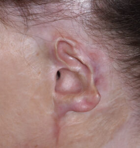
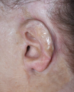 The healed result showed a reconstructed ear of normal height which provided a stable a good rest for her eyeglasses. She will require a second stage procedure to release the ear. from the side of the head fully with a skin graft to make a postauricular sulcus.
Case Highlights:
1) Burned ear deformities post difficult reconstructive challenges due to the loss of both cartilage and normal overlying and surrounding skin.
2) Usually the external auditory canal and the immediate surrounding cartilage framework is preserved.
3) Reconstruction of the outer ear framework requires rib cartilage covered by a temporal fascial flap and skin graft.
Dr. Barry Eppley
Indianapolis, Indiana