

Background: The contour of the inner leg below the knee is heavily influenced by the size and shape of the medical gastrocnemius muscle. This muscle takes it origin from the medial head of the femoral condyle and extends down the inner aspect of the lower leg to join with the soleus tendon/muscle on its way down to the achilles tendon. The superficial location of the muscle ensures that it is visible externally.
Like all muscles the medial gastrocnemius can be variable in its size. A prominent muscle creates a visible outer convexity which provides shape to the inner leg profile through its visible protrusion. A smaller muscle size may not create any convexity at all and leave the inner leg with a straight profile. Patients with this muscle size are typical candidates for implant augmentation.
But some inner leg deficiencies are not always due to muscle size. There are a few patients that have a very deep concavity right below the inner knee. The projection of the muscle may be adequate down further in the leg but it is inadequate right below the knee. Whether this represents a lower origin of the muscle off of the femoral condyle or lack of upper muscle thickness can be debated. But augmenting this area of the inner calf requires a different approach to implant placement and shape.
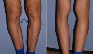
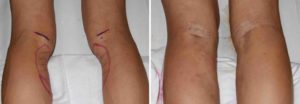
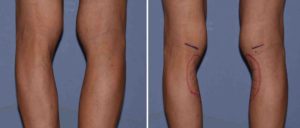 Case Study: This middle-aged female presented for inner calf augmentation. She was bothered by the severe indentation below her knee and the lack of calf ‘muscle’ fullness in this area. Calf implants were decided for augmentation but the actual zone of augmentation was higher and more medial than the location of the muscle belly.
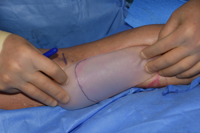
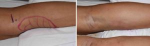
 Under general anesthesia and in the prone position, standard calf implants were used and could be seen to be much bigger than the needed zone of augmentation. These implants were carved down to the needed vertical height and feathered at their superior end. Then through a medial skin incision in a skin crease behind the knee, dissection was carried below the superficial fascia but on top of the deep fascia on top of the medial gastrocnemius muscle along the outline of the pocket location on the skin. The implants were then inserted and the superficial fascia, dermis and skin layers closed.
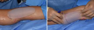
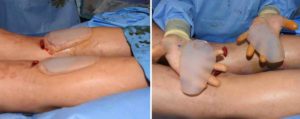
 The immediate intraoperative results shows a significant improvement in the profile of the inner leg with elimination of the deep concavity.
This unique area of inner calf augmentation unfortunately precludes subfascial placement of the implant. This always raises the question of whether any implant edge visibility will occur when all the swelling subsides and tissue contraction occurs around the implant over time.
Case Highlights:
1) The upper medial concavity below the inner knee is not a typical calf muscle deficiency.
2) A special designed ‘calf’ implant is needed for its correction that sits on top of the upper calf muscle fascia.
3) The upper medial calf implant sits higher than the traditional calf muscle implant.
Dr. Barry Eppley
Indianapolis, Indiana