

Background: While the most common chin procedure is augmentation a lesser number of chin shape changes are done for reduction. Besides their lower numbers chin reduction surgery is also dominated by female patients much more so than men. Chins that stick out too far, are too vertically long or are too wide are the dimensional changes that are bothersome for which different types of chin bone osteotomies and ostectomies exist to make these reductive chin changes.
Like all facial reshaping changes there is always the risk that the patient may regret hit later. No matter how much computer imaging is done beforehead and how convinced the patient is that they will be benefit from it, a small percent of patients can never adapt to the facial shape change. Sometimes it is because the patient ‘misses’ their old face and does not recognize the new one. Other times the facial shape changes have create issues that did not exist beforehand, particularly in the overlying soft tissues. These are typically problems such as soft tissue laxity or sag, adhesions and tissue stiffness.
Being a projecting structure the ability to reverse a chin reduction requires knowledge of how much chin bone was removed and how was it done.
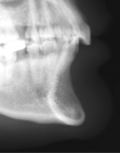
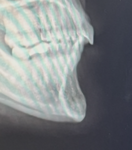 Case Study: This female had a bony chin reduction done one year previously. The reduction waS done by a burring technique through a submental incision. This was evident by a before and after lateral cephalometric x-ray that she had. It was estimated that up to 4mms of chin bone projection had been done. She did not like the blunted look of her chin, the skin dimpling that occurred over it as well as a new chin asymmetry on the right side.

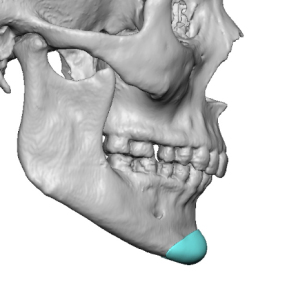
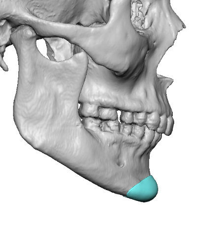
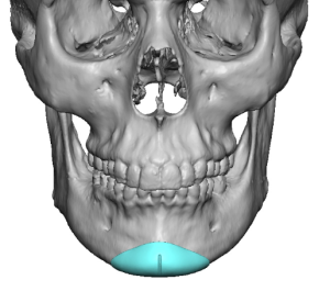 From a 3D CT scan a custom chin implant was designed that felt it most completely corrected the reduced chin point and corrected the asymmetry.
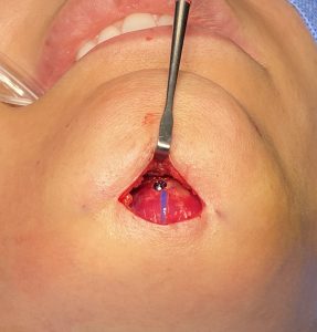 Through her existing submental incision the soft tissue chin pad was released from the bone where greater right side reduction was seen. The custom chin implant was modified slightly (reduced by 1mm on its sides) and she feared being slightly too big. The implant was inserted, positioned as designed and secured with a single microscrew.

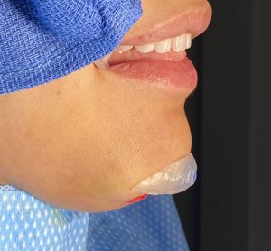
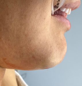 Her intraop profile showed the modest improvement in her chin projection.

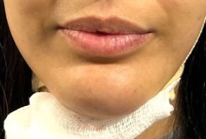 Her day after pictures from the front view showed the improvement in the chin shape with elimination of the dimpling.
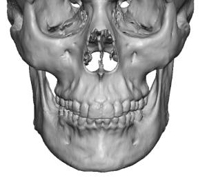
The most accurate method to reverse a chin bone reduction begins with a 3D CT scan. This provides the best assessment of what might have been reduced by seeing the actual shape of the chin. Reduced areas often look abnormal or with an irregular shape. While comparing a before and after 3D CT scan would be the ideal method to known exactly what was removed, few patients ever have had a before surgery scan.
Case Highlights:
1) Just like any facial reshaping procedure chin reduction can be a source of surgical regret with patients wanting to partially or full reverse the procedure.
2) For the accurate chin bone restoration a 3D CT scan is needed from a which a custom chin implant can be designed.
3) While there is not a method to know exactly how much chin bone was removed, the 3D shape of the chin and any prior pre surgical x-rays are helpful guides for this estimation.
Dr. Barry Eppley
Indianapolis, Indiana