


Background: Residual defects of the forehead and orbital regions are very common from prior early craniofacial surgery procedures. While moving skull and facial bones around at a young age are an ideal time for the bones to heal and open bone gaps to fit in, long term such skull and orbital bone reshaping procedures can still have residual partial and full-thickness bone defects.
While many of these bone contour defects can be filled in and covered over with bone cements as an older child or teenager, certain full-thickness defects can not. One of these craniofacial defects that requires a solid reconstruction is that of the lateral brow/orbital rims. This is one of the four walls of the orbit for which there is no substitute for an assured solid reconstruction. It is a rare craniofacial defect that is both complex in shape and must be solid as it supports numerous soft tissue attachments.
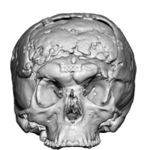
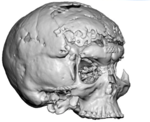
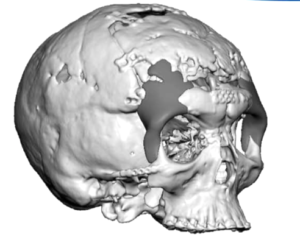
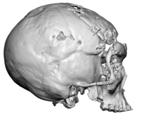 Case Study: This female patient had a prior history of numerous craniofacial procedures one of which was a hypertelorism repair. As she grew into an adult it was apparent that she lacked lateral eye support with down slanting eyelids and a very narrow lateral orbital/temporal width. A 3D CT scan showed the complete absence of the left lateral orbital rim and only a small remnant of a lateral orbital rim on the right side.
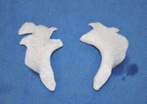
 Using the same scan custom implants were designed to create the missing lateral brow and orbital rims. The material, HTR (hard tissue replacement), which is made up of a hydrophilic porous PMMA/pHEMA was used to make the implants.
![]()
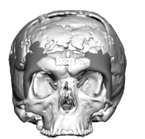
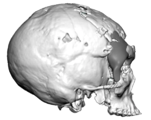
![]() A clear view printed skull model showed indwelling plates and screws in pink of which those around the lateral brow/orbital rim defects would need to be removed.
A clear view printed skull model showed indwelling plates and screws in pink of which those around the lateral brow/orbital rim defects would need to be removed.

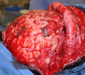
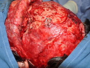 Under general anesthesia and with complete turndown of a coronal scalp/forehead flap through her existing scalp scar, many of the easily accessible indwelling plates and screws were removed. The lateral brow an orbital rim spaces were dissected out to expose the periorbital tissues particularly on the left side.
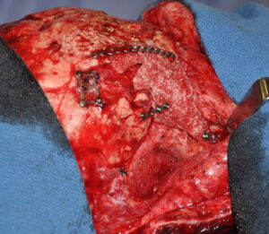
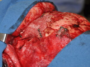
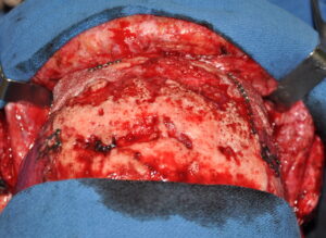 The custom HTR implants were soaked in an antibiotic solution and placed as per the design. 1.5mm plates and screws were used for implant fixation. The outer corner was then attached to the HTR lateral orbital rim to create canto support.
HTR implants has been around for three decades and has an excellent track record of use in full thickness skull defects. Its hydrophilic nature and intramaterial porosity allows for extensive tissue ingrowth. These material characteristics makes it superior to the use of autologous cranial bone grafts those survival in an open space (where much of the graft does not contact bone) is far from assured.
Case Highlights:
1) Complex full thickness bone defects of the brow and eye region requires a custom implant design approach.
2) Reconstruction of the important lateral brow and orbital rim defects require a synthetic material that allows tissue ingrowth.
3) The HTR material can be custom made into complex shapes and its rigidity requires requires a wide exposure for placement.
Dr. Barry Eppley
Indianapolis, Indiana