

Background: There is a natural alignment of the lower eyelid with eyeball for functional purposes that also has an aesthetic effect. Unlike the upper eyelid opens and closes the lower eyelid has less dynamic action. It largely is suspended from one side of the bony orbit to the other like a clothesline with its lid margin being up tight against the globe. With the lid against the eyeball, usually at a horizontal level or even with the outer corner slightly higher than the inner corner, the tear fluid used to keep the outer eyeball from drying out, flows towards the punctual hole located on the inner eyelid margin. The fluid then drains into the lacrimal sac from the puncture and eventually downstream into the nose.
The position of the lower lid is aesthetically judged by its position on the iris. Ideally the central lower lid covers 1 to 2mms of the colored iris. This is deemed to be a good aesthetic location. When it sits lowers than the edge of the iris more white of the eye is shown known as scleral show. While there are lots of people who naturally have scleral show due to the position of their lower eyelid it is judged as unaesthetic when int has become changed to either surgery or trauma. This becomes particularly bothersome when it has occurred in just one eye where the opposite lower eyelid sits higher resulting in noticeable eye asymmetry.
The usual lower eyelid asymmetry is when it sits lower due to scar contracture . The position of the eyeball is normal and it is the lower lid that its responsible for the scleral show. In rare cases of lower eyelid asymmetry the culprit is not the lower lid but that the eyeball is sitting too high. This can be due to an intraorbital pathology or from reconstruction of the orbital floor where the eyeball has been over elevated by too much augmentation of the orbital floor. In the reconstruction origin the most obvious solution is to lower the orbital folklore to drop down the eyeball to reduce the scleral show.
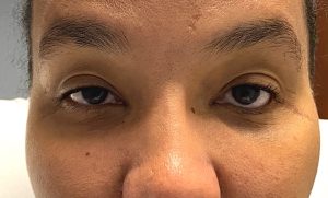 Case Study: This female sustained a severe left zygomaticomaxillary complex (ZMC) fracture and subsequently underwent multiple bony and soft tissue surgeries to rebuilt the cheekbone and orbital floor. Her remaining issue was some residual eye asymmetry where the left eye was higher than the non-injured right side with several millimeters of scleral show. Her CT showed that the orbital floor was covered by a mesh plate and bone grafts. She also had soft tissue cheek asymmetry where the upper cheek had too much fullness from prior fat injections and the undereye area was slightly hollow.
Her treatment plan consisted of microcannula liposuction of the left cheek and fat injections to the left undereye hollow done through a revised lateral canthal scar. The left lower eyelid was to be elevated by a full thickness skin graft.
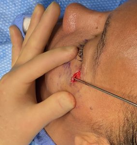
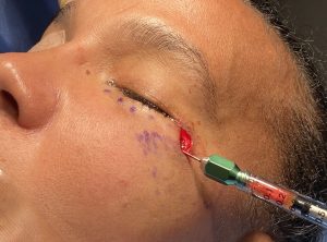 Initially the existing lateral canthal scar was excised. This provide access to first do fat reduction of the cheek through a 2mm three hole cannula. A surprising amount of fat was able to be removed from the prior fat injections. Then using 2ccs of abdominal fat harvested from inside the umbilicus the undereye areas was injected with a 1m cannula. The lateral canthal scar was then primarily closed with 6-0 plain sutures.
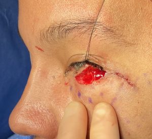
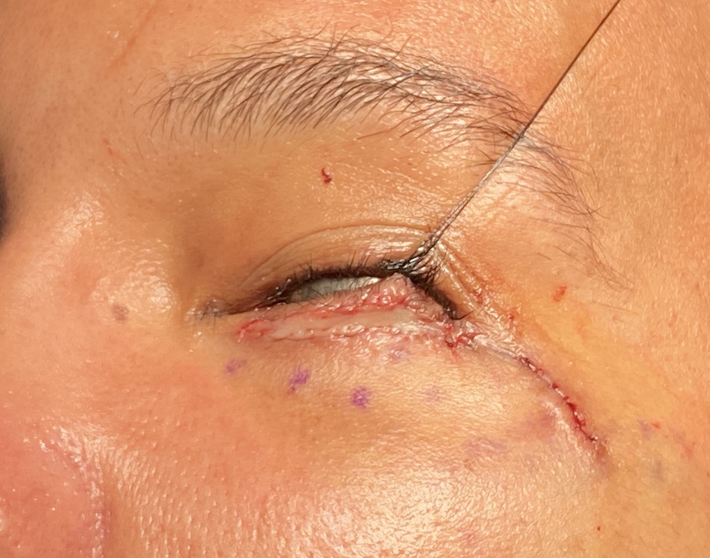
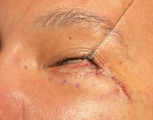 The left lower eyelid subciliary incision was open across the entire low margin and released with the aid of traction placed in a suture through the lash line. The orbicularis muscle was released and a full thickness skin graft harvested from behind the ear was placed into the newly created lower lid defect. It was sewn in with 6-0 plain and a thin xeroform bolster applied for compression by tie over resorbable sutures.
While her eye asymmetry was caused by a higher than normal globe position from her prior reconstructions, the effort that it would have taken to remove the mesh plate to lower the orbital floor did not seem to be a good choice. She would so close to the finish line that a more significant procedure for treating 2mms of scleral show was a poor risk:benefit ratio. Thus the unusual decision to raise the eyelid. Given her prior surgeries and the scar in the lower eyelid trying to raise the lid margin by spacer grafts alone I dod not think would work well. Placing a full thickness skin graft would be a more reliable technique by adding skin. There was going to be an expected color mismatch between the graft and the lower eyelid skin. But the small size of the graft would make that issue minimally perceptible.
Case Highlights:
1) In scleral show that results from an over elevated eyeball the effort and trauma to take apart the reconstructed orbital floor seems a solution that is bigger than the problem.
2) In this case it was better to raise the lower eyelid than try and lower the eyeball.
3) Raising the scarred lower eyelid is most reliably done by release and the placement of a full thickness skin graft.
Dr. Barry Eppley
World-Renowned Plastic Surgeon