

Background: The back of the head is typically perceived as being made up of the occipital skull bone. But this is not completely accurate. This large unpaired bone is the most posterior part of the skull and does makes up a significant part of its posterior wall. But the upper part of the back of the head is made up of the paired parietal bones which are separated from the occipital bone beneath it by the lambdoid suture lines.
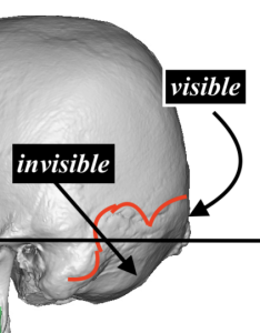 Below the lambdoid suture lines lies the visible occipital bone down to the occipital knob. But the invisible part, which represents its lower half, gives under the neck muscle and heads down to partially encircle the spinal cord. The relevance of this anatomy is that when an occipital bun deformity occurs the enlargement is only seen in the visible part from the central knob area superiorly. Interestingly most occipital buns do not have a prominent knob presumably because the bun protrusion has overgrown the knob area.
In occipital bun reductions the amount of bone removal possible is determined by the thickness of the outer cortical layer of bone. It can be reduced throughout the visible occipital bone down to the nuchal line/ridge. While a prominent occipital bun is always due to thicker bone and can always be visible reduced, it is usually prudent to get at least a preop lateral skull film to see how thick the bone layers are.
Case Study: This male had a prominent occipital bun which by x-ray showed a thick outer cortical bone layer which could be significantly reduced.
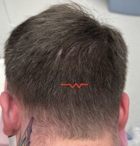
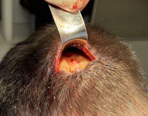 Under general anesthesia and in the prone position the location of the small irregular scalp incision located over the nuchal ridge line. Through this incision the bone color could be seen to have yellow tint and full thickness outer cortical cuts were made down into the marrow space in a radiating manner from the incision location.
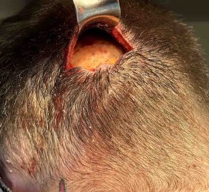 Using the depths established by the cortical cut lines the intervening bone was removed in a side to side manner, mindful of keeping a curved and not completely flat shape. The incision was closed over a drain.
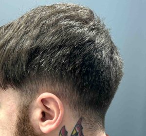
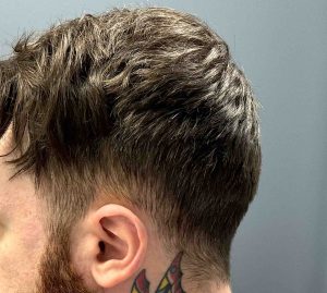 The drain and head dressing was removed the next day and the bun reduction could be appreciated.
Occipital bun reduction is done by removal of the visible outer cortical bone thickness. It will always make a difference in the amount of visible protrusion, it is just a question of how much.The occipital bone is the thickest of all the skull bones and the bun deformity represents an abnormal thickening of what is already a thick bone. A preoperative lateral skull film will show how thick and how much reductions can be achieved.
Key Points:
1) A prominent occipital bun is when a protrusion of the upper half of occipital bone exists
2) How much an occipital bun can be reduced depends on the thickness of the outer cortical bone layer.
3) In most occipital bun reductions a visible reduction is seen in profile but complete elimination of all of the bun’s prominence is rarely achieved.
Dr. Barry Eppley
World-Renowned Plastic Surgeon