

Background: Prominent temporal arteries are an aesthetic issue that is the direct result of an abnormally enlarged but normal structure. Every one has a temporal branch off of the carotid artery which ascends vertically into the hair-bearing temporal region and then splits into a Y configuration. (now called the superficial temporal artery or STA) At this Y the artery changes from a vertical into more of a horizontal orientation with anterior and posterior branches. The anterior branch heads out into the non-hair bearing area of the temporal and forehead areas. (anterior branch of the STA)
The anterior branch of the STA is what can become dilated (more prominent) and aesthetically bothersome. Since an accessory branch can also come off the temporal artery below the Y split and becomes dilated it is another source of a prominent temporal artery but it is distinguished by having a distinct vertical orientation as the artery ascends into the forehead. With these anatomic differences in the prominent temporal artery I have classified them into two types, type 1 is the result of the anterior branch of the STA while type 2 is the result of an enlarged accessory branch.
The typical type 1 prominent temporal artery requires 3 distinct ligation points. The most proximal is either right at the Y takeoff (in a shaved head male) or where it leaves the temporal hairline onto the open skin area. The most distal ligation point is at the visible end of the artery at the upper portion of the forehead before it goes into the scalp. The middle ligation, while to some many seem unneeded as the two ends have been tied off, is absolutely critical. This is done where the horizontal artery takes an abrupt turn northward at the lateral forehead aura. (what I call the elbow of the anterior branch of the STA)
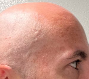
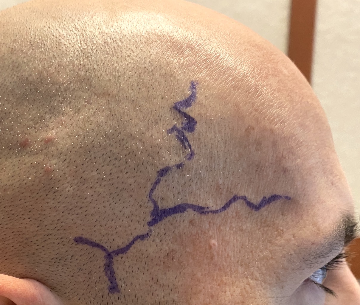
 Case Study: This male had prominent temporal arteries as long as he had shaved his head. With his shaved head the visible patterns of the temporal arteries at the Y and outward could be clearly seen. On the left side there was the typical type 1 pattern. On the right side there was, however, an additional Y split after the first Y out on the anterior branch.

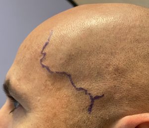 The arterial patterns between the right and left side is best seen once they are marked right before surgery.
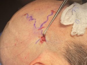
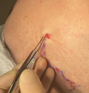 Under local anesthesia at each ligation site (4 on the right and 3 on the left) a small incision was made and the artery dissected out. They were double ligated with 5-0 prolene in the more proximal sites and 5-0 Vicryl at the more distal sites.

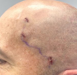 At the completion of all ligations a doppler was used to check for any arterial signals which were now silent.
Case Highlights:
1) Type 1 prominent temporal arteries come from the anterior branch of the superficial temporal artery and have more of a horizontal orientation.
2) Type 1 prominent temporal arteries typically require 3 ligation points for maximum reduction.
3) The double Y-branched type I prominent temporal artery is uncommon and requires an extra ligation point.
Dr. Barry Eppley
World-Renowned Plastic Surgeon