Background: Bony prominences of the skull can occur anywhere on its five surfaces in wide variety of shapes and sizes. But there are some consistent types of bony protrusions seen on each skull surface. These include sagittal crests on the top of the head, occipital buns and knobs on the back of the head, bossing and horns in the forehead and excessive temporal bone and muscle convexities on the side of the head.
Some of these prominences occur in or around suture lines which account for their occurrence. Such is the case of the sagittal crest which is the direct result of a thickening or hypertrophy of the midline sagittal suture line. On the back of the head the occipital bone may become prominent as it grows up against the bilateral inverted lambdoid suture lines.
Typically many bony skull prominences occur as solitary entities. But multi surface area involvements are possible and often there is an association between the two. Sometimes it is obvious by looking at how the skull develops as in the following case.
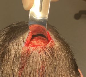
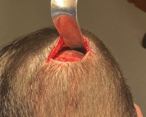
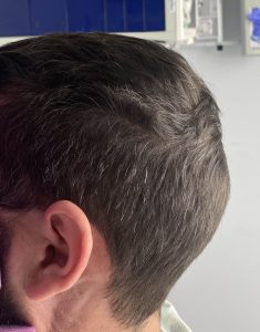
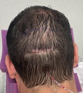
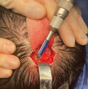
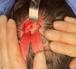
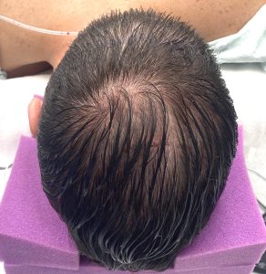
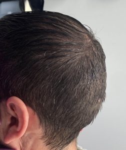
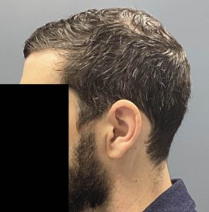
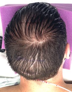
Case Study: This male was bothered by his head shape with two specific areas, a posterior sagittal crest and a prominent occipital bone with a ridge line up against the lambdoid suture line. One might have argued that the parietal areas above the occipital bone was deficient but there was a clear prominent occipital ridge.
Key Points:
1) Multi-surface skull reductions can be done during the same surgery and often involve separate incisions to do so.
2) Occipital skull protrusions are usually reduced through scalp incisions placed low on the bottom end of the bone over the nuchal ridge.
3) A posterior sagittal crest is reduced with a small scalp incision at its back end.
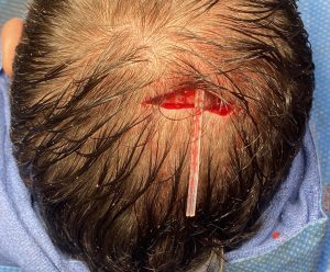
4) Drains are usually needed on many skull reductions and if they are in close proximity a single drain may work for both reduced areas.
Dr. Barry Eppley
World-Renowned Plastic Surgeon


