

Background: The occipital bone is one of eight cranial bones that make up the skull. It makes up the lower half of the visible back of head but what is seen is less than half of its entire size. The rest goes under the neck muscles and wraps around the vertebrae and spinal cord. Given its size it has been anatomically described as a giant vertebra that supports/cradles the brain. Because of its position and purpose it is also the thickest cranial bone.
The occipital bun deformity is a well known aesthetic skull protrusion that creates a distinct outcropping of the back of the head. While there are various head shapes that can create occipital elongation (e.g., scaphocephaly) the occipital bun is distinctly different as the skull shape around it, the parietal and temporal bones, are normal in development. Thus the occipital bun has a distinct outcropping of enlarged bone along the lambdoidal suture line which marks a clear contour deformity at its union with the paired parietal bones above it.
Because of its location and the attachments of the neck muscles along its nuchal lines the occipital bun deformity represents an enlargement of an already thicker skull bone. As a result significant reduction of its enlarged contour projection can usually be achieved.
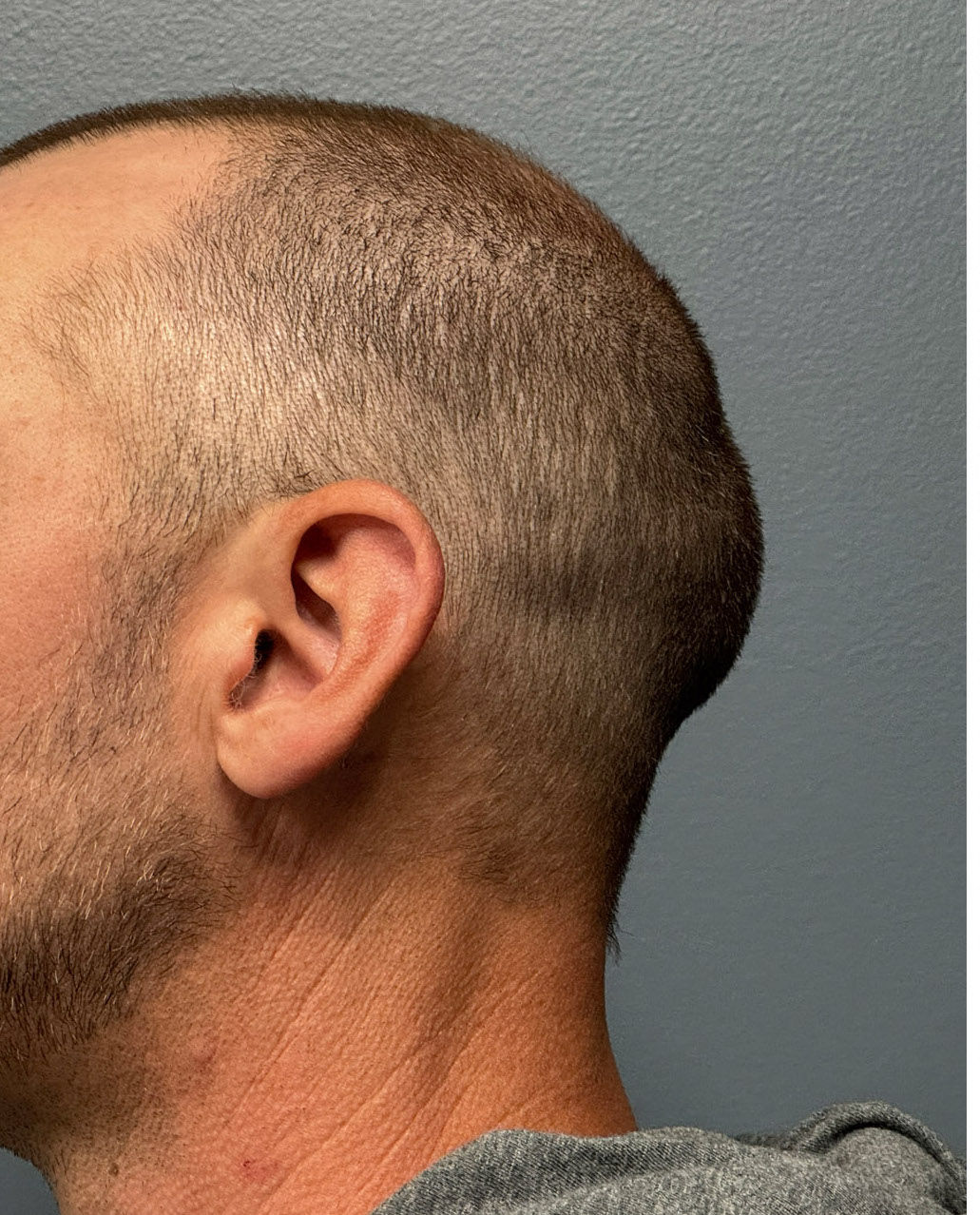
 Case Study: This male with a short haircut had a noticeable protrusion on the back of his head. Its enlargement below the lambdoidal suture line could almost be seen through his scalp. Computer imaging was done to show him the type of occipital bun reduction I thought could be achieved to make sure it was enough of a change for him to undergo surgery.
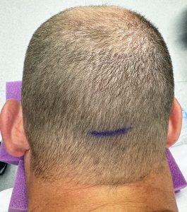
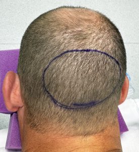 Under general anesthesia and in the prone position the occipital bone reduction was performed through a small horizontal incision over the nuchal ridge. The extent of the occipital bone enlargement was marked in a circular pattern.
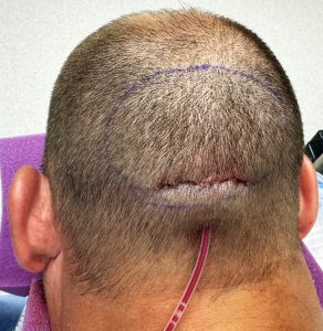 Using a high speed handpiece and round carbide burr (7mms in thickness) the outer cortex of the enlarged occipital bone was reduced down to the diploic space. In doing the reduction it is important to maintain the curved shape and not make a flat surface. Closure of the incision was done with small dissolvable sutures over a drain.
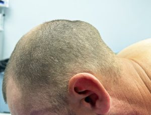
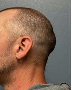
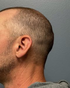
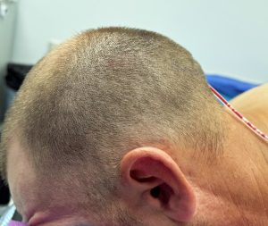 In looking at the side profile of the back of his head on the operative table in the prone position, the reductive change appeared consistent with what the preoperative imaging suggested was possible.
The drain was removed the next day along with the head dressing on his own.
The occipital bun skull deformity is always because of thicker bone development as evidenced by its protrusion above the lambdoidal suture line. The question is not whether its prominence can be reduced but by how much. (completely or incompletely) A preoperative x ray can help make that determination but the reduction can usually be done down to the level of the lambdoidal suture line and the parietal bone above it.
Key Points:
1) The occipital bones of the skull is the most common overly grown/developed bone that creates an undesired aesthetic protrusion. (occipital bun)
2) The occipital bone is located at the bottom half of the back of the head of which its superior portion is the only visible part.
3) Most occipital bun protrusions can either be partially or completed removed for a satisfactory back of the head reshaping.
Dr. Barry Eppley
World-Renowned Plastic Surgeon