


Background: The ear is composed of a complex cartilage shape that accounts for much of its external appearance. The simplest shape of the cartilage, the auriculocephalic angle, happens to be the one that is the most requested ear shape change and the most commonly performed aesthetic surgery. (setback otoplasty)
The auriculocephalic angle is defined by how much the ear sticks out from the side of the head. This has been classically defined as normal if the angle is in the range of 25 to 35 degrees. While this is an anthropometrically determined number what really counts is how the patient determines their ear position and what looks good to them. Most patients have no knowledge about their actual auriculocephalic angle measurement. What they do perceive is whether the helical rim is visible from the front view or not.
Inability to see the helical rim from the front view most commonly occurs from otoplasty surgery. Some patients who have had the surgery would view their result as overdone if the helical rim is pulled back behind the antihelical fold. This serves as the basis for otoplasty reversal surgery which really should be called subtotal otoplasty reversal surgery since complete reversal is not desired. The goal is to bring back out the helical rim a few millimeters into visibility usually at the center portion of the ear.
But in very rare cases the ear may naturally have developed a tighter auriculocephalic angle when the helical rim is not visible at all. This means that the ear cartilage and overlying skin is naturally tighter. Making the helical rim more visible in these cases poses some challenges not seen in the more traditional subtotal otoplasty reversal after a setback otoplasty.
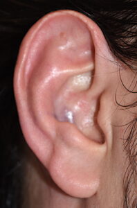
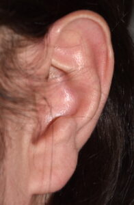
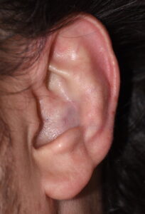
Case Study: This male, who had never had any ear surgery, wanted the middle operation of his ears to stick out further. The natural shape of his ears showed a helical rim that had less projection than that of the more inferior earlobe.
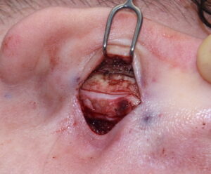
 Under local anesthesia a small incision was made on the back of the ear over the mid-concha. With cartilage exposure a series of vertical releasing cuts were made in the conchae cartilage to allow it to open up the auriculocephalic angle. The expanded concha was support outward by carving a piece of cadaveric rib cartilage to serve as a vertical strut support between the concha and the mastoid fascia in the depth of the postauricular sulcus. It was sutured with through and through sutures between the rib cartilage and the conceal cartilage.
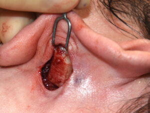
 Both ears incisions were closed and the ear position inspected. Both the patient and I agreed that even more projection was desired. To try and achieve that change a small incision was made behind the helical rim and the antihelical fold cartilage released. An additional cartilage graft was placed into the fold to help hold it out, effectively creating a t-shaped cartilage support to hold the ear out even further.

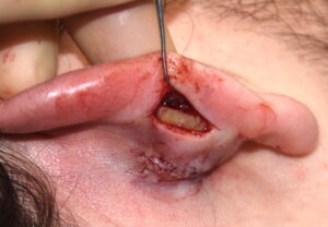
 Once these incisions were closed it as felt this was the maximum amount of ear protrusion that could be achieved.
The reverse otoplasty in a naturally tighter ear position along the side of the head is much more challenging to change than the traditional over corrected setback otoplasty. Unlike releasing ear cartilages that have the required amount of cartilage for expansion, significant new cartilage is needed to push out the ‘cartilage deficient’ decreased auriculocephalic angled ear. One postoperative outcome the patient should thus expected is that the ear will feel very stiff for a prolonged postoperative time period. This is no different that in a rhinoplasty where a columellar strut has been added to the tip of the nose. The natural suppleness of the nasal tip will be lost for a long time as it heals and may never completely feel as supple as it once did. The ideal issue applies to that if the ear. The very cartilage support needed to increase the projection of the ease will make it very stiff for some postoperative time.
Case Highlights:
1) The most common requested change in the auriculocephalic angle of the ears is to lessen it, making it wider or more open is a rare request unless it is after a setback otoplasty.
2) Increasing the natural auriculocephalic angle is to usually achieve a more visible helical rim that is visible beyond the anti helical fold in the front view.
3) Pulling the ear further from the side of the head requires ear cartilage releases with cartilage grafting to support it outward.
Dr. Barry Eppley
Indianapolis, Indiana