

Background: The occipital bone is associated with the back of the head and is generally believed that is the primary bone of it. In reality it makes up only a portion of the back of the head and is only located at the visible lower portion. When one realizes that the majority of the occipital bone envelopes the spinal cord (foramen magnum) and given where the bone is positioned on the skull, one can envision it as a giant vertebrae that supports the brain. Only the upper portion of the occipital bone is what is seen from an aesthetic skull shape standpoint.
When the occipital bone becomes enlarged a specifically shaped protrusion occurs on the back of the head that sticks out abnormally. This is typically seen as an oblong or football-shaped protrusion that is both palpable and visible even if one has hair. In many cases the occipital protrusion is due to an abnormal thickening of the bone beneath the lambdoidal sutures. But it can also be normal thickness bone that is pushed out by the growth of the bone. (this is what is seen in sagittal craniosynostosis)
Reduction of the prominent occipital bone, often call the occipital bun, can be done by a bone burring technique. The entire outer layer of cortical bone can be removed right down to the diploic space. This is usually enough to make a visible external change that creates a more even contour of the back of the head. The hardest part is being to do so through a fairly limited scalp incision.
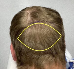 Case Study: This male was bothered by a football-shaped protrusion on the back of his head that he was noticeable to him after puberty. Given that he wanted to wear a shorter haircut, he desired it to be reduced.
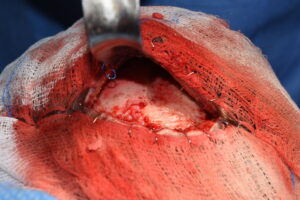
 Under general anesthesia in the prone position and through a low horizontal scalp incision (the hair protected by sponges stapled to the incision), an initial midline vertical bone cut was made down through the protrusion using a high speed handpiece and burr. Once the depth of the bone reduction was established the right half of the bony protrusion was reduced and contoured. The left side of the protrusion was then similarly reduced and contoured. Any persistent bone bleeders were covered by bone wax.
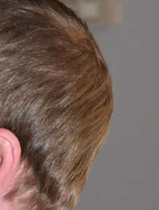
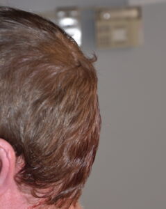
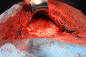
The immediate before and after improvement in the shape of the back of the head could be seen at end of surgery.
 Partial or complete reduction of occipital bone protrusions can be safely and effectively done through a low scalp incision. While checking a CT scan before surgery is not routinely done, it can provide information as to how much bone can be shaved down to the inner cortical layer. When viewed in a sagittal section the line of reduction could be made to determine if an adequate reduction can be achieved.
Case Highlights:
1) The occipital bone makes up only a portion of the visible back of the head
2) Occipital bony protrusions occur up to the intersection of the sagittal and lambdoidal sutures with an oblong shape.
3) Removal of the outer cortical layer of the occipital bony protrusion (skull reduction) creates a visible change in the shape of the back of the head.
Dr. Barry Eppley
Indianapolis, Indiana