

Background: The skull is composed of a number of plates of bone which are joined by sutures. These sutures allow the skull to grow with the underlying developing brain. Once brain growth is complete these sutures became stiffer and more fibrotic and even partially calcified. In adults the major suture lines are still seen of which the most visible are the midline sagittal and the transverse coronal and lambdoidal sutures. The sagittal suture is the most recognized one due to its midline location on the top of the head.
The sagittal suture is unique amongst the cranial sutures in my experience because it can be become abnormally thick and raised creating a ridge or crest. No other cranial suture in adults exhibits this topographic shape. This makes the vertex (highest point of the sagittal suture) very visible and gives the head a peaked shape as seen in the frontal view. Such sagittal crests often exhibit no visible suture line indicating a hyperostotic response, suggesting a minor expression of the craniosynostosis spectrum.
In reducing the aesthetic sagittal crest deformity the bone can be reduced by high speed burring. Because the sagittal crest bone is always thicker than that of a normal adult suture line its can usually be reduced enough to make a visible external difference. This eliminates the peaked midline point in the frontal view and reduces the height of the vertex in profile. The key is achieving that result through the smallest scalp incision as possible.
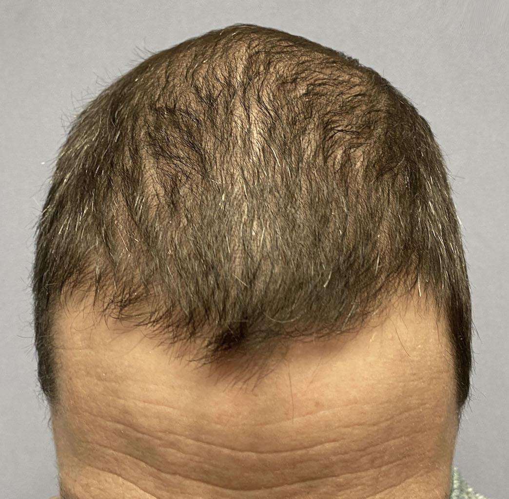
Case Study: This male was bothered by a midline crest on the top of his head. He had an overall more narrow head which magnified the presence of the crest. With very short hair he was called ‘Bone Hawk’ at work, presumable because of the resemblance to the shape of the hawk’s skull.
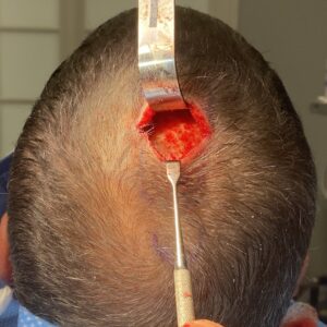
 Under general anesthesia a small 3cm scalp incision was made transversely over the midpoint of the length of the crest. (11cms in length) Subperiosteal elevation was done in both directions allowing for a bony reduction to be done throughout its entire length. Using a high speed guarded burr the outer cortex of the crestal bone was reduced. The deepest reduction was done at the level of the incision down next to the diploid space. Scalp closure was done with resorbable sutures over a drain that traversed the length of the reduced bony crest.

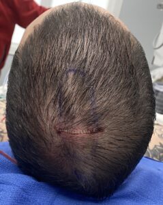
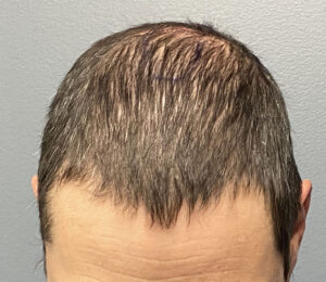 When seen the next day for head dressing and drain removal the improvement in the shape of his head could be seen.
Sagittal crest skull deformities are the result of a thickening of the suture between its anterior coronal suture intersection and its posterior end at the original posterior fontanelle location at the crown. While it can have thickening anywhere along its length it is usually located more posteriorly. In shorter sagittal crests (7cms or less) the scalp incision is placed on the back end of they crest. In longer crest reductions the incision needs to be made at the midpoint of its length so equal access can be obtained to its entire length. The tunnel technique allows for only limited exposure and working length.
.Case Highlights:
1) The posterior sagittal crest skull deformity creates a visible peak at the top of the back of the head best seen in the frontal view.
2) Reduction of the sagittal crest is done by high speed bone burring down to the level of the diploic space.
3) A tunnel technique allows the midline bone reduction to be done through the smallest possible scalp incision.
Dr. Barry Eppley
World-Renowned Plastic Surgeon