

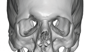
Background: While the brow bone is often perceived as a uniform raised bar of bone above the eyes, its topographic anatomy is more complex. The medial brow bone is made up of the expanded underlying frontal sinuses with a midline dip between them due to the septum which separates the paired sinus cavities. The outer half of the brow bone is composed of solid frontal bone. What separates the inner and outer brow bone is the supraorbital notch or bony exit of the supraorbital nerve.
Trauma affects the brow bones differently dependent upon whether it occurs on the medial or lateral parts of the bone. Impacts on the medial brow bones usually results in comminuted fractures and inward displacement of the anterior wall of the frontal sinus. Conversely impacts at the tail of the brow bone often result in inward and downward displacement of the bony rim with fracture lines through the supraorbital notch medially and the frontozygomatuc suture line laterally.
When brow bone fractures are not treated initially, secondary reconstructions can be done to recontour the malaligned bony rim.
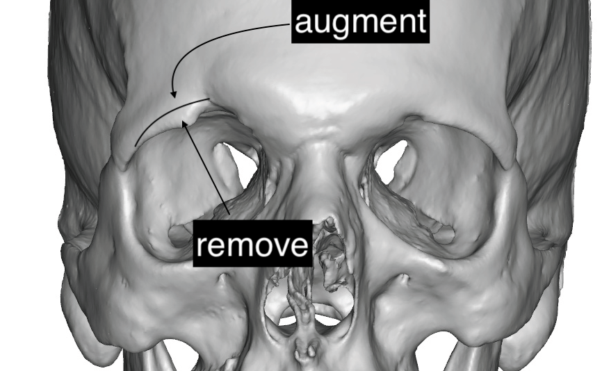
 Case Study: This male had a prior history of a traumatic injury to the right lower forehead resulting in a fracture with downward and inward displacement of the lateral brow bone causing increased fullness of the upper eyelid. A 3D CT scan confirmed the malposition of the tail of the brow bone with a healed fracture line/indentation through the supraorbital notch.
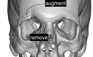 Surgical correction was to be done by removing the lower edge of the brow bone and an onlay augmentation of its flatter anterior surface.
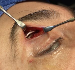
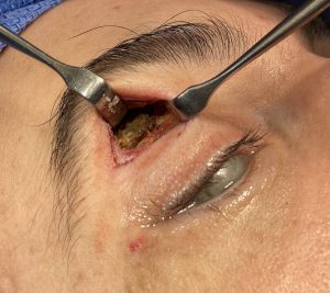 Through a small upper eyelid incision the lower edge of the brow bone was removed using a high speed handpiece and a small shaping burr.
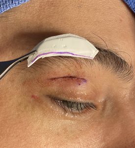
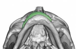
 A 2mm thick piece of ePTFE sheeting was shaped to fit the brow bone and onlayed onto its surface. It was sutured to the periosteum during closure.
While the medial half of the brow bone can only be accessed through a superior approach, the lateral or tail of the brow can be approached either from superior (scalp) or inferior (eyelid) directions. For many isolated tail of the brow bone reshaping needs the upper eyelid incision provides more direct access. It is also the most convenient method when the inferior bony border needs to be raised…which is much more difficult to do from a frontal hairline or scalp incisional approach.
Case Highlights:
1) Lateral brow bone reshaping can be done through an upper eyelid or transpalpebral incision.
2) Through an upper eyelid incision the lower edge of a lateral brow bone can be most reliably reshaped vertically.
3) Moderate amounts of lateral brow bone augmentation can be done by ePTFE sheeting or bone cements through the upper eyelid incision.
Dr. Barry Eppley
Indianapolis, Indiana