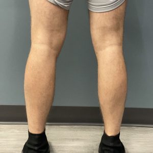
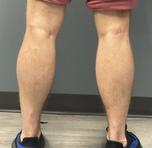
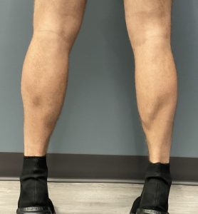
Background: The calf muscles, known as the gastrocs, are some of the hardest muscles to build up by exercise. They are so stubborn to enlargement because they are already working regularly by just the walking and standing that we normally do. Thus to get them even bigger than they normally are they have to really be challenged. It is not that they can’t be made bigger by exercise it is just that being harder to do than other muscles people often give up with lack of immediate or easy results.
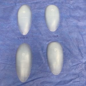
But if one decides to forego weight training and go for an immediate and dramatic appearance of larger calf muscles, the surgical placement of calf implants can achieve that effect. When placing calf implants the initial decision is whether to undergo a 2 or 4 implant augmentation. The most important calf muscle from an aesthetic standpoint is the inner or medial muscle. This is the larger of the paired calf muscles and is the one seen from the front view. Whether this muscle alone should be augmented or both muscles implanted depends on the patient’s objectives. If improvement of skinny lower legs is the goal the inner muscle alone can just be done since this improves their appearance between the knee and ankle from the front. But if the goal is an enhanced muscular appearance then both calf muscles of each lower leg should be augmented.
Sizing the calf implants for each patient is done by measurements of the muscle size (length and width) and then choosing the implant dimensions that are just a bit smaller. Since the calf implant will the placed in the subfascial location and the overlying fascia is tight a slightly smaller implant than the muscle size is prudent.
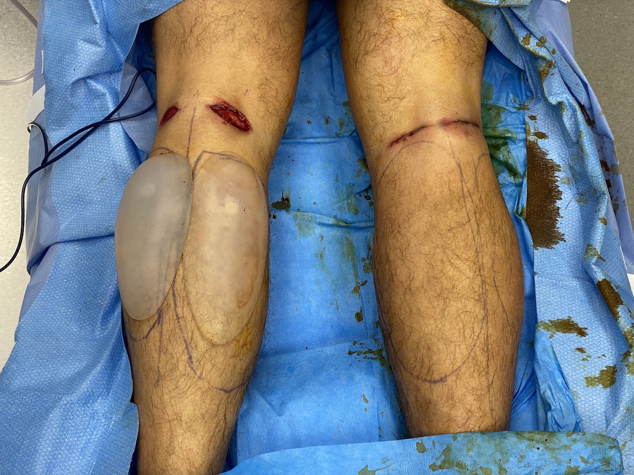
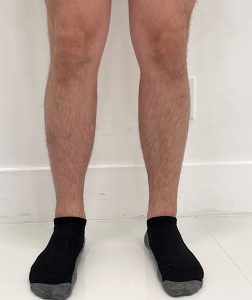
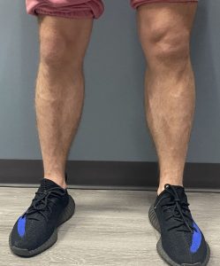
Case Study: This male had a well developed upper body but not get the legs to match, particularly the lower legs despite his best efforts. He desired enlargement of both inner and outer bellies of the gastrocnemius muscles.
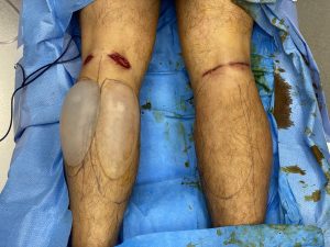
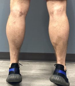
Calf implants can be a fairly straightforward aesthetic augmentation procedure of the legs. There are two nerves to avoid in their placement. The sensory sural nerve runs within the fascia between the two calf muscles and can be avoided by keeping the inner and outer calf muscle pockets from communicating. (hence the two separate incisions. The lateral peroneal motor nerve is at the the outside of the outer muscle pocket and can occasionally be seen during the dissection down to the fascia as it wraps around the fibular head. It is best avoided by not making the outer skin incision too lateral.
There is one last issue to be aware which is unique to the placement of bilateral calf implants. Large calf implants in a tight small leg has the potential of causing a compression syndrome due to vascular compression. I have never seen it but it pays to be aware that it could happen and checking capillary refill in the toes and/or nailed oxygen saturations levels postop is prudent.
Case Highlights:
1) Lower leg augmentation can be successfully done by the placement of calf implants in the subfascial pocket.
2) The decision in calf implants is whether they should augment just the larger inner calf muscle or both the inner and outer calf muscles.
3) In placing calf implants it is important to know the location of the sural (sensory) and lateral peroneal (motor) nerves.
Dr. Barry Eppley
Indianapolis, Indiana

