

Background: The temporomandibular joint (TMJ) creates the articulation with the only moveable bone of the face. (lower jaw) It is responsible for allowing one to be able to open their mouth whose extent is most evident in the distance between the upper and lower front teeth. (interincisal distance) Like all joints it is composed of a ball (mandibular condyle) and socket (glenoid fossa of the temporal bone) but it has the additional element of a fibrocartilaginous disc or meniscus between them.
While often perceived as a simple hinge joint, how it actually works is more complex…and it has to do the disc inside the joint space. The hinge function of the joint occurs when the lower jaw initially opens due to rotation of the condyle inside the joint. This may be an interincisal distance of up to 10mms. But what makes the lower jaw really open is translation. This is where the condyle comes out of the fossa (dislocates itself) which is made possible by the diametric movement of the smooth disc. This allows the interincisal opening to become as great as 35mm to 50mms.
TMJ ankylosis occurs when the lower jaw is unable to open due to an obstruction within (intracapsular) or around the joint. (extracapsular) Either can be a source to prevent jaw movement. There are a wide variety of causes of TMJ ankylosis and trauma is a major factor in younger patients where facial growth is very active.
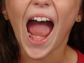
 Case Study: This female child presented with a diminished ability to open her mouth one year after sustaining a subcondylar fracture after falling from her bike. She had good centric occlusion with a 9mm opening with a deviation towards the originally injured side. A preoperative x-rays showed a healed subcondylar fracture with some bend to the condylar neck. The joint space was radiopaque.
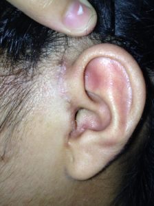
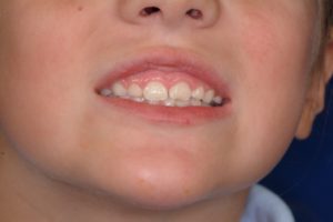
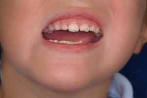
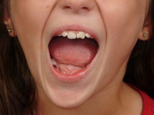 Under general anesthesia and through a preauricular incision the TMJ space was released and a small fat graft placed. Her interincisal opening at six months after surgery was 45mm with a persistent slight right side deviation.
TMJ ankylosis may be caused by fibrosis or actual bony union. The former is far more common.When surgically released two issues must be considered. What is going to prevent recurrent scarring and motion limitations (hence the fat graft) and wha type of after surgery physical therapy will be needed. In children the simple use of stacked tongue blades are a convenient and inexpensive source of increasing jaw motion.
Case Highlights:
1) TMJ ankylosis can be caused by intracapsular or extracapsular factors.
2) A preauricular incisional approach is needed for intracapsular TMJ release.
3) Postoperative physical therapy is needed to ensure that some of the interincisal distance achieved during surgery is maintained months after surgery.
Dr. Barry Eppley
Indianapolis, Indiana