


Skull reduction is a specialized cranial contouring surgery designed to reduce and reshape areas of the skull to create a smaller, smoother, and more proportionate head shape. The procedure involves thinning or removing portions of the outer skull bone to achieve the desired contour.
It is most commonly performed for aesthetic purposes—to refine a high, wide, or asymmetric skull—or as part of gender affirmation surgery (facial feminization) to reduce the skull’s height or width and soften cranial contours. It may also be indicated for reconstructive correction of asymmetries, trauma-related deformities, or irregularities from prior surgeries.
Common Skull Areas Addressed
-
Top of the head – to reduce height or flatten a peaked skull.
-
Back of the head (occiput) – to lessen protrusion or correct asymmetry.
-
Sides (parietal regions) – to narrow skull width or reduce bulging above the ears.
-
Forehead – when included as part of overall facial balancing or brow bone contouring.
Surgical Technique
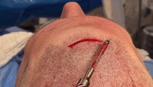 The only effective method for reducing bony skull areas is burring (bone shaving). A high-speed burr gently removes the outer table of bone to smooth or reduce projection. The key to success lies in understanding how thick the skull is and how much bone can be safely removed.
General Principles of Skull Bone Thickness
Skull thickness varies significantly by region, sex, and age. General adult averages are as follows:
|
Region |
Typical Thickness |
|---|---|
|
Frontal bone |
6–8 mm (up to 10–12 mm in hyperostotic males) |
|
Temporal bone |
2–4 mm (thinnest in the squamous portion) |
|
Parietal bone |
5–7 mm |
|
Occipital bone |
8–15 mm (thickest at the inion) |
Additional variables:
-
Sex: Males typically have thicker skulls overall.
-
Age: Thickness increases slightly until middle age, then may decrease with advanced aging.
-
Ethnicity: Documented population differences exist (e.g., thicker skulls in African populations vs. thinner in Caucasian populations).
While these are reliable general guidelines—and consistent with my clinical experience from hundreds of skull reduction cases—modern technology now allows a far more precise preoperative assessment. Using plain x-rays or even 2D CT scan assessments, while useful in their day, have been supplanted by more advanced bone thickness assessment techniques.
Contemporary Imaging and Assessment Techniques
Computed Tomography (CT) is the gold standard for skull thickness evaluation.
Recommended CT Parameters:
-
High-resolution, submillimeter slices (?1 mm)
-
Bone window settings (W: 2000, L: 400)
-
3D reconstruction for direct thickness mapping and surgical planning
MRI is not suitable for this purpose, as it lacks adequate bone signal.
Measurement Methods
Manual Measurement
-
-
Use calipers or measurement tools in DICOM-viewing software (e.g., RadiAnt, 3D Slicer, Mimics).
-
Measure perpendicularly from the outer to the inner table over the intended reduction area.
-
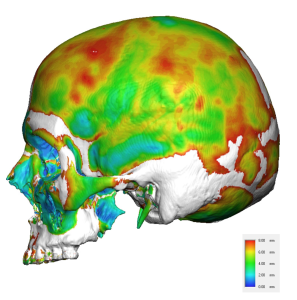
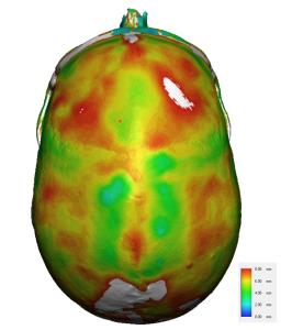
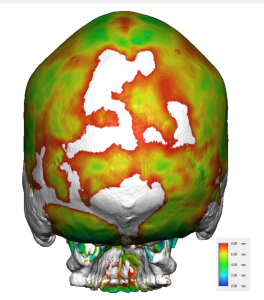
Color Mapping (Perferred Technique)
-
-
Segment bone using threshold-based algorithms.
-
 Generate color-coded maps displaying local bone thickness.
 -
Allows comprehensive visualization and quantitative assessment of the entire skull.
-
 Generate color-coded maps displaying local bone thickness.
3D Skull Model Segmentation
– 3D skull model is created
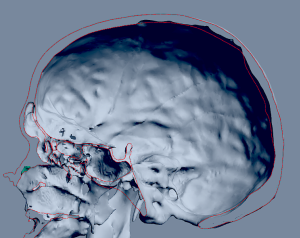
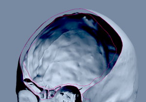
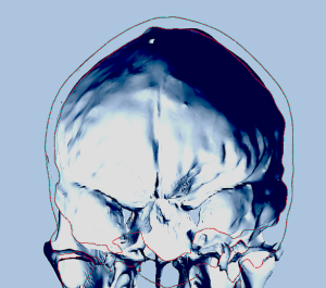 – individual skull bone slices can be taken from any orientation and bone thickness measured
The central question in planning skull reduction is not whether the procedure can be performed—it can—but whether it will be effective in achieving the patient’s aesthetic goals.
Quantifying how much bone can be safely reduced provides the most valuable preoperative insight for both surgeon and patient.
Preoperative Imaging Protocol:
-
Acquire a thin-slice craniofacial CT scan (?1 mm slice thickness).
-
Load DICOM data into 3D modeling software (e.g., Mimics, Materialise, 3D Slicer).
-
Segment the bone and generate a 3D skull model.
-
Apply a thickness analysis tool to visualize and quantify bone thickness.
-
Correlate imaging findings with the surgical contouring plan.
Dr. Barry Eppley
World-Renowned Plastic Surgeon