

The ear is composed of two basic structures, cartilage and skin. The cartilage component of the ear is considerable as only the earlobe is not supported by it. The cartilage is solely responsible for the very complex shape of the ear with its many hills, valleys, ridges and curves that are seen externally. How it gets this shape is an embryological marvel as six hillocks fuse in utero to ultimately create what we see as the external ear.
While cartilage supports all the convexities and concavities of the ear, its most important contribution is to its elevations or convexities. (helical rim, superior and inferior crus, antihelix, tragus and antitragus) Cartilage can be removed from any of the concave areas and the shape of the ear would not change. This is well known from the common harvesting of ear cartilage in rhinoplasty from the concha, the largest ear concavity which looks the same both before and after graft harvest.
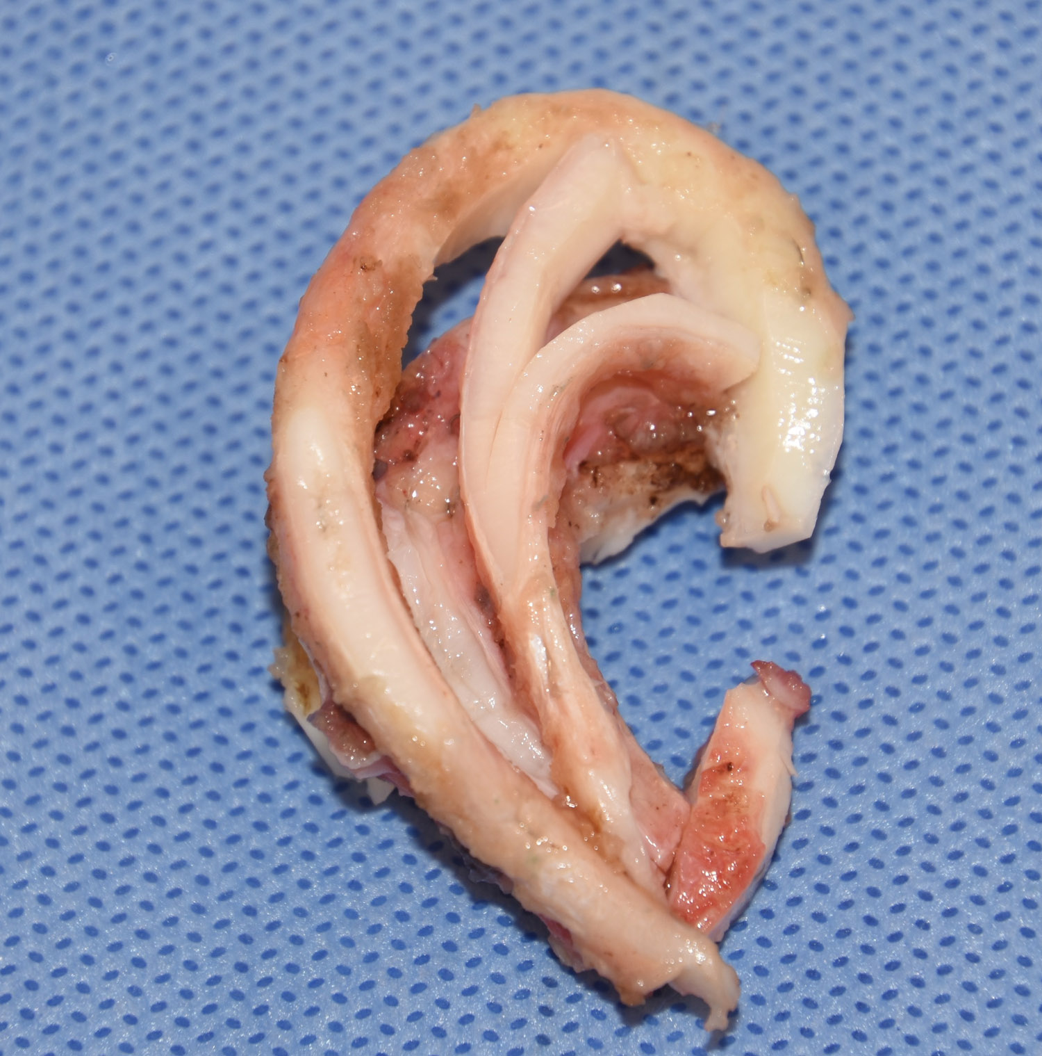 The greatest illustration of the role of cartilage in the shape of the ear is in microtia reconstruction. For children born with parts or all of the external ear missing, the traditional ear reconstruction method is done with rib cartilage. Portions of ribs 6, 7, and 8 are used to create a cartilage ear framework for insertion under the skin. In making his ear framework the complete concept of the ‘hills and valleys’ of the ear must be artistically created by carving and assembling the pieces of rib cartilage. The eventual shape of the ear is seen many months after surgery as the overlying skin shrinks into and around its cartilage shape.
Of all the shaping procedures that are done in plastic surgery throughout the body, making an ear out of rib cartilage in microtia reconstruction certainly qualifies as a sculpting surgery.
Dr. Barry Eppley
Indianapolis, Indiana