


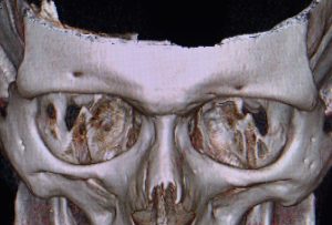
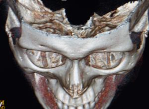
 Brow bone augmentation is one of the least performed facial augmentation procedures as evidenced by the lack of any preformed implant to do it. This does not mean, however, that male patients don’t have brow bone deficiency concerns and a desire to address it. One uncommon indication for brow bone augmentation is asymmetry where one brow bone has less projection than the other side or is at a different vertical level. This is most evident by looking at the different locations of the supraorbital nerve foramen which otherwise would be even on both sides.
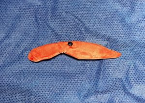 The ideal method to treat a brow bone asymmetry which involves a deficiency is to make a custom brow bone implant by computer design to match the other side. The alternative non-custom implant approach is to use 2mm thick ePTFE sheeting which can be hand cut to the general size of the defect and the edges crimped for feathering. Most brow bone asymmetries are usually no more than a few millimeters in deficient projection. To keep the brow bone implant low enough on the brow ridge, as the soft tissues have recoil and tend to push it back up, a single screw fixation is most ideal.
There are two approaches to placing a limited in size brow bone implant, superiorly using an endoscopic approach and inferiorly through the upper eyelid. (transpalpebral) If the implant only covers lateral to the supraorbital nerve then the upper eyelid approach can accomplish good placement in a more direct manner. Bur if the implant must cross over the supraorbital nerve and go medial to it then the endoscopic approach will allow more assured positioning with nerve protection and the ability to still use screw fixation.
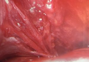
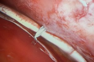 Using a single port access through a one cm vertical incision right behind the hairline the subperiosteal pocket is created, visualizing the nerve as usual in the process. The brow bone implant is inserted longitudinally and then turned horizontally to slide onto over the brow bone. The supraorbital nerve will be right below the implant with the notch cut out for it in the implant. The best technique for placing a single fixation screw is using an 18 gauge needle passed percutaneously through the eyebrow. This will show the location of the skin entrance site to the implant.
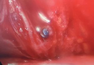
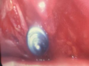
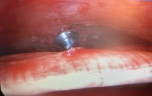
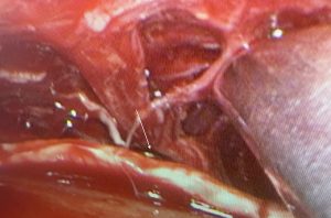 Then a short self-tapping 1.5mm screw on the driver is passed right through where the needle was initially placed in the eyebrow. It can be seen endscopically passing between the branches of the supraorbital nerve, through the implant and then driven flush with the implant’ surface.
The challenge in any brow bone implant is placing it with minimal scarring and ensuring that it stays in place. Endoscopic visualization with percutaneous screw fixation can effectively achieve these brow bone implant goals.
Dr. Barry Eppley
Indianapolis, Indiana