


Background
The sagittal crest is a raised midline ridge of bone most commonly seen in many mammals and reptiles. It serves as the attachment site for the temporal muscles—developmentally indicating strong jaw function. In general, the higher the sagittal crest, the larger and stronger the temporal jaw muscles (e.g., gorillas, certain dinosaurs).
Humans typically do not have a sagittal crest in normal skull development. Over evolutionary time, the origin of the temporal muscles has shifted more to the sides of the skull, producing a rounder cranial shape. This adaptation may have occurred to accommodate an increased brain size. Aesthetically, even a hint of a sagittal crest is generally not considered desirable.
In humans, pronounced sagittal crests can occur due to congenital craniosynostosis—specifically premature fusion of the sagittal suture. More commonly, smaller crests arise from localized bony enlargement along the suture line without actual synostosis. The cause is not well understood, but they are often seen in individuals with sloped parasagittal areas and a more narrow head shape.
Case Study
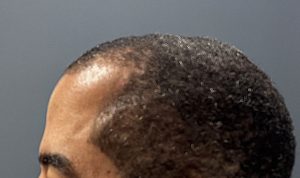
 This male patient had long been self-conscious about a midline hump at the front portion of the top of his head. In profile, this created a neutral or slightly reversed cranial slope, with the anterior and posterior heights of the skull nearly level. On palpation, a distinct bony step-off was felt leading onto the back portion of the raised ridge.
Surgical Technique:
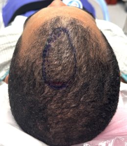 Under general anesthesia, the sagittal crest was marked and a small incision made at its posterior end. The crest was exposed, revealing a visible suture line.
-
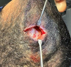
Exposure & Preparation: Wide subperiosteal undermining was performed well beyond the crest to create adequate working space.
-
 Bone Reduction: Using a high-speed handpiece and burr, the crest was reduced, revealing increased bone thickness (as expected). Both sagittal and coronal suture lines were identified, indicating the crest began at their junction and extended forward toward the frontal hairline.
 -
Closure: After maximal safe reduction, the scalp was closed over a drain.
 Bone Reduction: Using a high-speed handpiece and burr, the crest was reduced, revealing increased bone thickness (as expected). Both sagittal and coronal suture lines were identified, indicating the crest began at their junction and extended forward toward the frontal hairline.
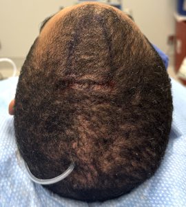 Closure: After maximal safe reduction, the scalp was closed over a drain.
Postoperative Result:
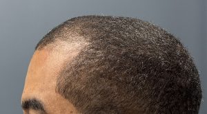
 The following day, after dressing and drain removal, the improvement in cranial slope was clearly visible in profile, with a smoother anterior contour.
Discussion
Sagittal crests most commonly occur posteriorly, between the coronal suture and the anterior edge of the original posterior fontanelle. This location typically produces an exaggerated cranial slope with a higher posterior skull. Less commonly—as in this case—the crest lies anterior to the coronal suture. In such cases, the deformity may relate more to the metopic suture than to the sagittal suture. Anterior sagittal crests are often accompanied by a narrow upper forehead, consistent with lateral growth restriction and compensatory superior midline growth.
Regardless of location, surgical feasibility depends on bone thickness. In every sagittal crest reduction performed by the author, the bone has been thicker than the surrounding skull, allowing for satisfactory contouring. If bone thickness is uncertain, a preoperative CT scan provides definitive assessment.
Key Points
-
The sagittal crest is an enlarged midline bony ridge often associated with a narrow head shape.
-
Anterior sagittal crests can create a raised or slightly reversed cranial slope in profile.
-
Surgical reduction typically reveals a fibrotic suture line beneath the crest and yields significant improvement in contour.
Dr. Barry Eppley
World-Renowned Plastic Surgeon