Custom design facial implants offer the opportunity to produce the most significant changes to the skeletal structure of the face. While not true in every case, many times these implants are far bigger and cover more bony surface area than that of standard facial implants.
By virtue of being larger they can be more challenging to surgically place and the margin of error in placement is actually much smaller. In a larger implant a slight change in tilt or positioning on the bone will lead to a more visible external facial asymmetry or shape change due its size. This is most dramatically illustrated in the custom jawline implant which is the biggest implant that can be made for the face covering one jaw angle to the other.
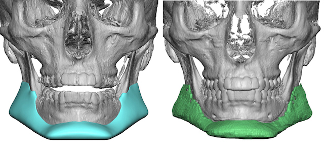
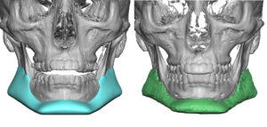
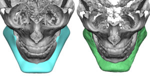
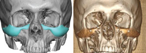
When their are facial asymmetry concerns in custom facial implants well after the swelling has resolved (6 to 12 weeks), an assessment of why requires a 3D CT scan. There is no accurate method by looking on the outside to really know and one certainly doesn’t want to undergo a revisional adjustment by ‘eyeballing’ it. If the asymmetry couldn’t be detected during its initial intraoperative placement by external inspection on the operative table then it is unlikely one would be more successful trying the same approach later. The following case examples illustrate the value of a postoperative 3D CT scan in cases of questions custom facial implant asymmetry.
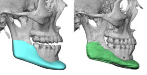
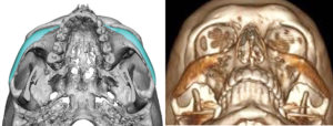
While both of these cases failed to demonstrate implant placement as the source of their asymmetry concerns, it shows the value of the scan in postoperative asymmetry concerns. More times than not there will be a detectable implant asymmetry which does account for what the patient sees and provides a clear understanding for the surgeon for subsequent implant revision.
It is also important to note, as can be seen in these examples, that the custom facial implant is little less distinct than it is on the design scan. This is expected as the design scan is a computerized drawing on the bone. This is different that a postop scan which is a computerized reconstruction of the imaged implant data.
Dr. Barry Eppley
Indianapolis, Indiana

