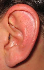
Background: The shape of the ear, like the nose, is one of the most variable features of one’s face. While it is chocked full of hills and valleys composed of cartilage and has an array of anatomic convolutions, the human ear is nonetheless very recognizeable. Any gross abnormalities in its shape and size is easily observed.

Known by a multitude of names such as dumbo ears or elephant ears, these very names indicate that it is not viewed as a favorable facial feature. While it may provide a better eyeglass resting place or a more effective method of holding one’s ear back, the social stigma that comes with prominent ears overrides whatever functional benefit that it may provide.
Surgery for the protruding ears, most commonly called ear pinning (there is no pins used in the surgery however), has been around for over 100 years. When it was originally introduced long ago, only skin was removed from the back of the ear to pull it back. This did not work well and it was eventually recognized that the actual shape of the cartilage needed to be changed to produce a better and more permanent result. Many otoplasty cartilage techniques have evolved over the years but the use of permanent sutures to make the fold remains as a main component of the operation.
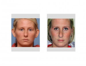
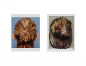
Case Study: This 26 year-old female had long been bothered by her protruding ears and finally decided to have them reshaped. She had a 65 degree auriculocephalic angle and lack of an antihelical fold cartilage prominence. Her ears were also very stiff and did not fold back very easily.


Otoplasty surgery for the protruding ears is incredibly effective and produces a dramatic change in the shape of the ear. How far the ear should be respositioned back along side of the head is a matter of intraoperative judgement.Overcorrection and undercorrection is always a concern but the most likely reason for revisional otoplasty surgery is asymmetry. Getting both ears in the indentical position with the cartilage reshaping is challenging and is alspo affected by how well the sutures hold and how the ears heal.
Case Highlights:
1) Ears that stick out are because the shape of ear cartilage is not adequately folded onto itself.
2) Otoplasty or ear reshaping is done by creating a fold in the main cartilage of the ear to bring back the outer helix closer to the side of the head.
3) The most common complication with otoplasty surgery is asymmetry which may require revisional surgery.
Dr. Barry Eppley
Indianapolis, Indiana


