Background: One of the most challenging of all plastic surgery procedures of the face is ear reconstruction. Microtia, or congenital absence of the external, is the most extreme challenge when it comes to making an ear essentially from scratch. A more common, although not less challenging, is that of partial ear reconstruction.
Whether it be from an automobile accident, a sharp edged instrument or even a bite wound, the loosely attached and flexible cartilage of the ear and its attached skin is relatively easy to avulse or be amputated. Loss of part or all of the ear cartilage, while not life-threatening, is nonetheless disfiguring and very psychologically disturbing.
I have seen ear amputations present numerous times with the avulsed ear segment in hand (or someone else’s hand) with the understandable hope that it may be able to be reattached. This is rarely possible although there is no harm in making an attempt if it is not overly crushed or mutilated, which it often is. Unless the ear is in fairly pristeen condition, it is better to close the wound and let it heal for six months before embarking on ear reconstruction efforts.
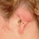
Six months later she presented for ear reconstruction. Initially tissue expansion was planned as the first stage prior to rib graft placement but she needed to complete her ear reconstruction as possible. (she was enrolling the military)
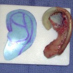
In her definitive one-stage ear reconstruction, the first step is to make a template of the opposite ear and transpose it to the amputated ear site to get the right shape and orientation of the reconstruction.
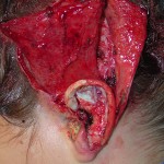
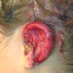
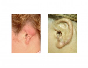
Case Highlights:
1) External ear reconstruction from partial or total amputation injuries is best done with rib cartilage when significant portions of the framework are missing.
2) Due to scarring and skin loss, more skin has to be created to produce an ample and supple skin cover over the rib graft. This is most commonly and easily done with tissue expanders which makes it a staged reconstruction approach.
3) A one-stage ear reconstruction approach can be done using a temporoparietal fascial flap and split-thickness skin graft coverage.
Dr. Barry Eppley
Indianapolis, Indiana


