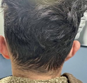
Background: Whether it is laying in utero or after birth the back of the head is exposed to the greatest duration of external forces or pressure. As a result of this external force exposure the back of the head has several anatomic features that distinguish it from other head area. First the occipital bone is the thickest of all the skull bones often being well over 1cm between the outer and inner cortices. Secondly the overlying scalp is also thicker than any other area on the head. Lastly, the insertion of the neck muscles at the bottom of the occipital bone assures it has multiple raised bony areas to support their ligamentous attachment further contributing to the thickness of the bone.
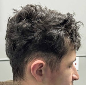
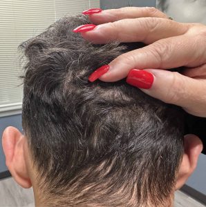
The shape of the skull is prone to deformations during development of which the back of the head is the most common area affected. The thickness of the bone or scalp does not resist these deforming forces which can lead to a variety of degrees of flatness (lack of projection) and asymmetries. While lack of projection and asymmetries of the back of the head can occur separately they are also quote common to occur concurrently.
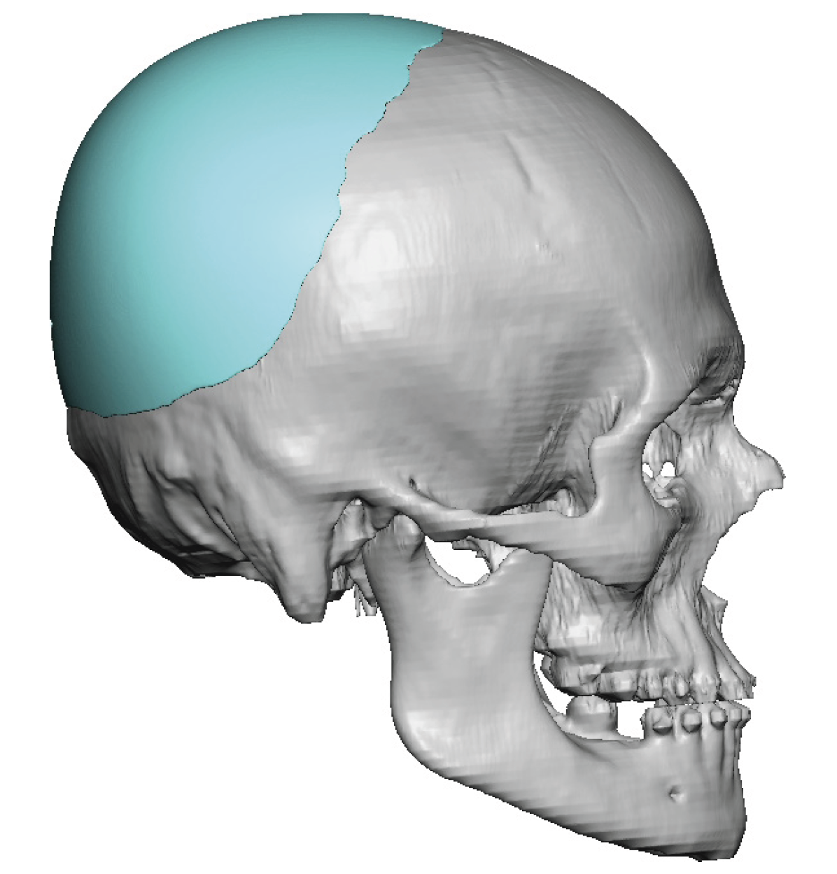
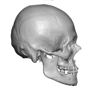
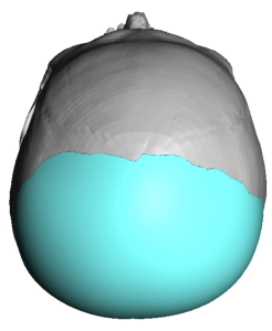
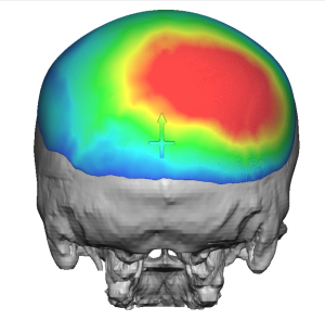
When correcting the shape of the back of the head due to asymmetric flatness there is only one effective way to do so….a computer implant design. It is impossible to adequately create an asymmetry correction an by eyeballing it alone. Not to mention getting a good projection shape that adequately covers the surface area of bony involvement. A 3D CT skull scan provides a full visual assessment of the scope of the deformity for all angles from which a good corrective design can be done.
Key Points:
1) It is not uncommon to have both a flat and asymmetric back of the head shape.
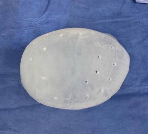
2) The computer design of a custom skull implant can accommodate for the differences in the shape of the back of the head and make it more symmetric.
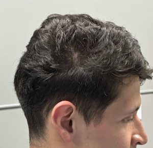
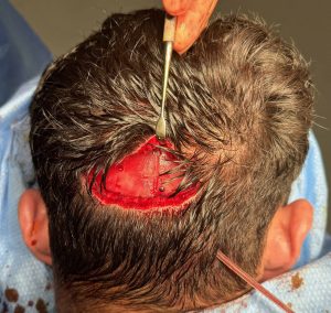
3) The back of the head custom skull implant is placed through a small scalp incision in which no hair is shaved to do so.
Dr. Barry Eppley
World-Renowned Plastic Surgeon

