


 The natural ear is a complex structure consisting of various convexities and concavities. The convexities are the outer helical rim and the Y-shaped superior and inferior crus and antoihelix. The concavities are the large inner most concha around the external auditory canal and the triangular and scaphoid fossas. While the functional significance of these combined ear shape geometries can be debated, their presence is essential in creating an acceptable looking ear.
Reconstruction of the ear is a well known surgical technique that uses either natural rib cartilage, an alloplastic implant system or a combination of both. From an implant use standpoint the key to a successful outcome is adequate and viable overlying soft tissue coverage. In some cases of congenital microtia adequate skin coverage may exist after the existing deformed and small cartilage framework is removed. If in doubt transfer of a vascularized fascial flap is needed. In adults in reconstruction of traumatic or oncologic ear structure loss local rotational skin flaps may be adequate. But the key is understanding that, unlike rib cartilage, a implant framework has little tolerance for compromised overlying skin whether it be due to tissue quality or lack of tension free skin coverage.
 The advantages of an ear implant framework is that it provides an immediate off-the-shelf reconstruction that removes the need for time consuming rib cartilage carving for the surgeon as well as a painful harvest site and a permanent scar for the patient. The advantage of an ePTFE-coated silicone ear framework is that is allows soft tissue adhesion due to its micrifibrillar surface and has the softness and flexibiity that is more consistent with that of the normal cartilage-based ear.
1. Preoperative Implant Selection and Preparation
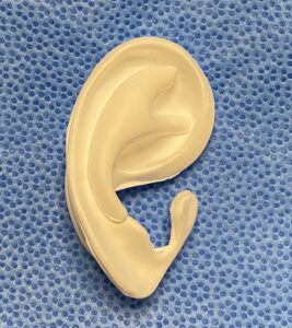
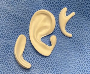 The ePTFE-coated silicone ear framework has three components, a base framework, a posterior block and an antihelix overlay. The base framework is the fundamental to which the posterior block is almost always used with it. The antihekix overlay is used only when additional enhancement of the framework is needed.augmentation is needed.
 The base framework comes in four sizes that range from 55mm to 63mms in height. This is the key framework dimension when selecting it for the patient based on the size of the opposite ear or the size of the ear the patient wants. The other dimensions of the framework (e.g., width) are controlled by the vertical height.
 The posterior block or wedge is used to push out the framework for improved ear projection (auriculocephalic angle) either during initial placement or as a secondary procedure. The block comes in four sizes rom 1.2 to 1.4cms of wedge push or projection. It fits behind the framework in a near interlocking fashion. The block can also be used to help push out a natural ear or rib cartilage framework where harvesting cartilage to do so is not desired.
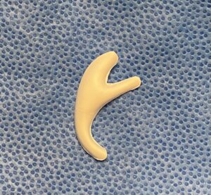 The antihelix overlay helps enhance the appearance of the aesthetically important Y-shape area of the upper part of the ear inside the helical rim. (superior and inferior crus = raised ridges, triangular fossa = concavity between the two crus) It can be used to add to the base framework during initial placement or as a secondary augmentation or can even be used to add to an existing rib graft ear reconstruction.
2. Intraoperative Handling and Implant Modification
 Made of ePTFE-coated silicone, Implantech’s ear implants are soft and flexible and can be easily cut or shaped if needed. The material has a consistency that is very similar to actual rib cartilage and can be modified with a scalpel in a similar fashion. The most common framework modification, in an effort to optimize the postoperative appearance of the details of the ear reconstruction, is to cut out various concave areas of the framework. This will allow the overlying soft tissue to contract into these framework areas.
The implants, once opened and prepared with any framework modifications, should be placed into a container containing an antibiotic or antibacterial solution until ready for implantation.
3. Implant Fixation and Closure
Fixation of the different parts of the ear implant can be done with sutures. ePTFE-silicone provides an easier material to suture together if needed than rib cartilage or other plastic implant materials. Such suturing is typically done between the base framework and the postauricular block. The antihelix overlay, if used, does not typically require suture fixation due to its Y-shaped fit.
The composite ear implant can be sutured to the underlying fascia on which it rests as needed to maintain implant position and angulation to the side of the head.
Prior to closure a drain is placed under the implant to aid in postoperative adaptation of the overlying skin into the details for the framework. After irrigating out with an antibacterial solution a two layer suture closure is then done over the implant. A lightly compressive ear dressing is applied being careful to avoid too much pressure and causing vascular compromise of the overlying skin.
Dr. Barry Eppley
World-Renowned Plastic Surgeon