

Background: The calf muscles, also known as the gastrocnemius muscles, makes up the back part of the leg between the knee and ankle. It is a two-headed muscle with an inner (medial) and outer (lateral) head that extends downward to form a common Achilles tendon which attaches onto the back part of the heel bone. (calcaneus)
Because of the stout tendinous attachment at the heel bone, Achilles tendon injuries ultimately affect the calf muscles as well. Through either inactivity (recovering from surgical tendon repair) and/or a change in tendon length between the end of the muscle and the bone, calf muscle deficits are well known to occur both in the short and long term. In many Achilles rupture patients full calf muscle strength and size may never fully recover despite maximal rehabilitation efforts.
When the origin of calf asymmetry is traumatic-induced muscle atrophy, the aesthetic appearance of it is often more significant than concerns about muscle weakness. Rebuilding calf volume can be done by either injection fat grafting or calf implants. Each has their advantages and disadvantages. The main advantage of implants is their assured permanent volume effects.
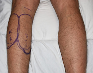
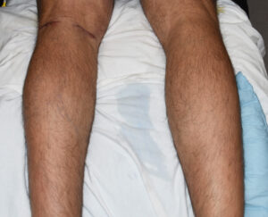
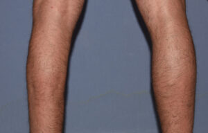 Case Study: This male had a history of an Achilles tendon rupture from a sport-related injury of his left leg. Between surgery and rehabilitation he remained with a persistent calf muscle deficit several years later. He opted for inner and outer calf implants in trying to create a better match to the uninjured right calf muscles.
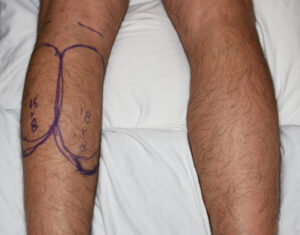 In selecting calf implants the pertinent issues are surface area coverage and volume. In asymmetry the normal side calf muscles lengths and widths are measured in extension. (toe stands) The calf implants that come closest to these measurements are chosen. The volume needed can not be precisely known and thus the only question is whether the implant projections will need to be thinned down or not. This will have to be an intraoperative decision.
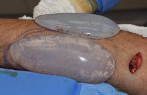 Under general anesthesia in the prone position, back of the knee skin incisions were made to create subfascial pockets over the medial and lateral calf muscles. The implants were inserted into the pockets and checked to the opposite leg for size comparison. No reduction of the implant volumes was needed.
 In using implants in calf muscle atrophy the volume needed for optimal symmetry correction can never be precisely known beforehand. The length and width of the muscles can be but not the volume. Interestingly I have always found it to be bigger than what one would initially think. So choose the implant that matches the calf muscle footprint and adjust its thickness during surgery if so needed.
Case Highlights:
1) Achilles tendon injuries can result in some permanent loss of calf muscle size.
2) Calf asymmetry due to muscle atrophy can be improved using standard calf implants.
3) Since both heads of the calf muscle will undergo atrophy due to a common cause, medial and lateral calf implants are often needed.
Dr. Barry Eppley
Indianapolis, Indiana