

Background: When significant atrophy of the deltoid muscle occurs a bony shoulder appearance results. This most commonly occurs after an axillary nerve injury. The significant shrinkage of the muscle skeletonizes the AC joint and the upper humerus creating significant shoulder asymmetry.
Restoration of the atrophic deltoid muscle can be done by soft tissue techniques such as fat grafting and muscle flaps. But fat injections have unpredictable volume retention and may require numerous fat grafting procedures to get a sustained visible result. A muscle transfer can be done from the latissimus dorsi muscle but between the harvest scar and the inevitable muscle atrophy that will come from a muscle transfer that lacks function stress on the fibers this is not an effective treatment option.
Implants offers a permanent volume retention method for significant deltoid muscle atrophy. It is also a favorable implantation site as it is a muscle bed even if much of the muscle mass has been lost. Determining the size and shape of the implants is the challenge and there are multiple methods to do so. 3D MRI imaging is the most accurate method to measure the exact muscle volume compared to the normal shoulder from which an implant design can be generated…but it is also the most expensive. The historic moulage technique is the most economical but not as exacting in terms of soft tissue volume loss.
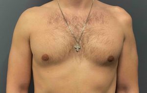
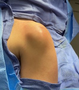
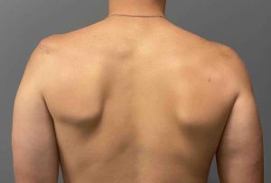 Case Study: This young male had custom deltoid implants for atrophy secondary to an axillary nerve injury from orthopedic shoulder surgery. These implants were made using a moulage technique. The results were a major improvement but he desired further volume addition.
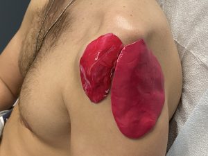
Using the existing two custom deltoid implant computer designs, the same footprints were maintained but their volumes increased. The original combined implant volumes was 68ccs. The new deltoid implants were 138cc, a 100% volumetric increase. A smaller 3rd implant was also designed to fill in a small hollow in front of the anterior deltoid muscle.
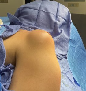
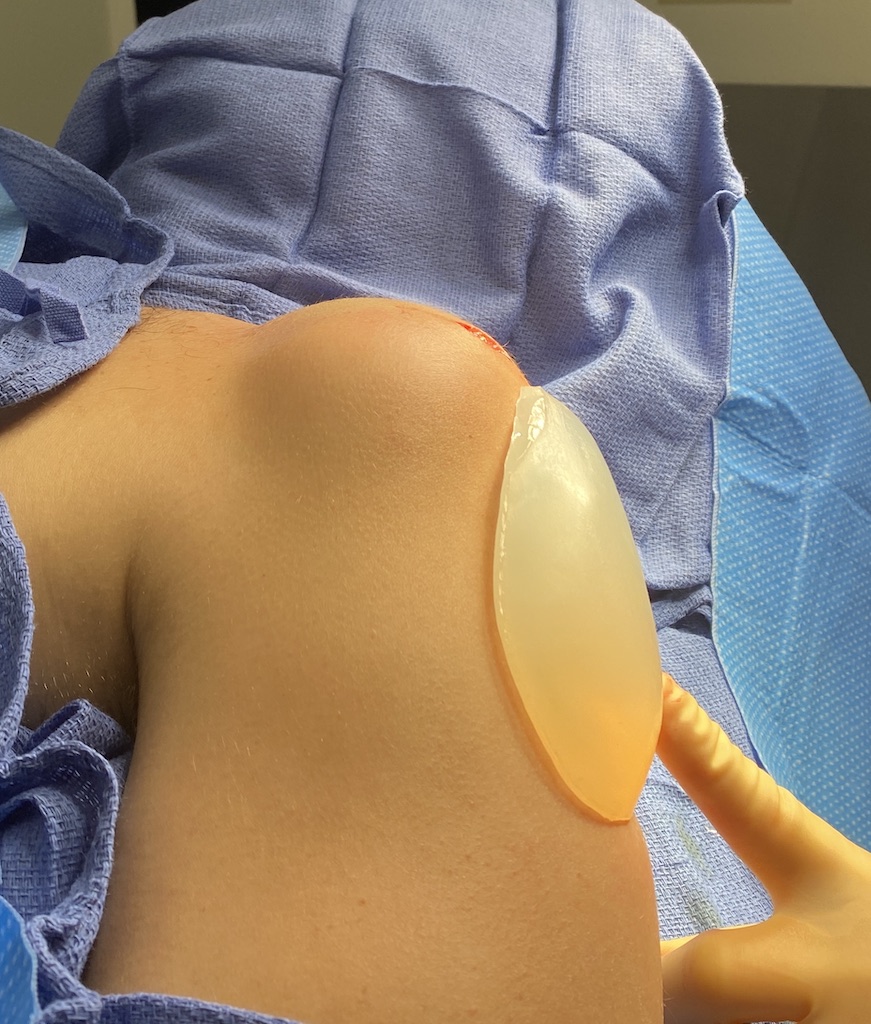
 Under general anesthesia and through his original two shoulder incisions the first set of deltoid implants was removed…revealing the original extent of the shoulder shape deformity. The larger deltoid implant, filling in for the central and posterior muscle heads, was then placed.
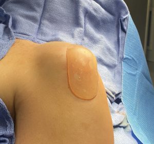
 The new more anterior deltoid implant was placed next.
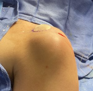
Lastly the very small implant, which went into a hollow at the upper pectoralis muscle, was inserted through the anterior incision.
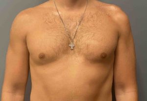
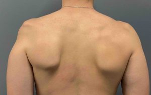
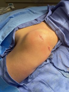 His result after this second set of deltoid implants showed a completely normal appearing shoulder with minimal scarring to do so.
 The moulage body implant fabrication method relies on the application of a quick-setting two silicone elastomer mixture onto the external contour deformity. It is then shaped until it appears to the eye to be the volume needed. Such an implant fabrication method is not an exact science and may over- or under estimate the implant volume needed. In the case of initial implant undercorrection the original implant designs serve as a guide on how to improve the original result.
Case Highlights:
1) Augmentation or restoration of the shoulder shape after deltoid muscle atrophy from an axillary nerve injury is most effectively done with custom implants.
2) The shape of deltoid implants in shoulder shape restoration is most conveniently done using a moulage technique in which the models are then scanned into a computer design.
3) If the initial deltoid implants do not provide optimal shoulder shape symmetry, new deltoid implants can be designed by further increases in volume from the 1st implant designs.
Dr. Barry Eppley
Indianapolis, Indiana