


 Background: Microtia is a well known congenital ear deformity that has established techniques for its reconstruction. The completely autologous method using rib cartilage grafts, typically done around age 6, is a proven technique that offers complication free long term results. The aesthetic success of the procedure is based on how well carved and shaped the rib graft framework is, its placement on the side of the head and how well the overlying soft tissues adapt down into and around it.
But one aspect of microtia rib graft surgery in children that is less discussed is how well the ear reconstruction grows/looks over time decades later. Does the reconstructed ear grow as the contralateral normal ear does or does it remain the same size as when it was placed? It is believed that the ear reconstruction does grow and thus it has been an established practice to match in size the ear rib graft framework to that of the normal ear. At the least it is recommended not to make the ear reconstruction in children an adult size in the belief it does not grow and the other ear will catch up in size to it eventually.
If long term the ear reconstruction ends up bigger than the other ear and is a noticeable aesthetic distraction as a result, how to secondarily shape the rib graft framework has not been reported to my knowledge. (although it most certainly has occurred)
 Case Study: This 40ish year old female was born with a left hemifacial microsomia and underwent a 3 stage ear reconstruction using rib cartilage as a child. (1st stage framework insertion, 2nd stage lobule rotation and tragus creation and 3rd stage postauricular sulcus release with skin graft) The major problem with it was how vertically long it was at 72mm in height with a 62mm in height opposite normal ear. There was also little definition of the upper 1/3 of the reconstruction and the conchal shape was round and disproportionately large.
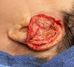
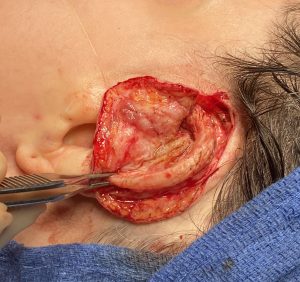
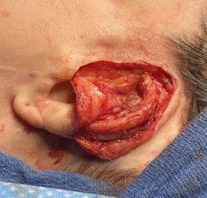 Under general anesthesia an incision was initially made along the entire upper helical rim and the upper cartilage framework exposed.The helical rim cartilage graft was separated from the underlying framework and rotated down in a more inferior position on the framework. It was stabilized there with permanent sutures. This cartilage maneuver shortened the height of the ear and increased the projection of the outer helical rim. The outer skin flap was thinned, excess skin graft removed and the upper cartilage framework recovered.
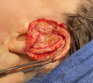
 Cartilages grafts were taken from the now exposed upper edge of the framework and used to create an antitragal prominence and a more visible superior helical root. Through and through resorbable sutures were placed to adapt the overlying skin flap to the cartilage framework and eliminate any dead space underneath it.
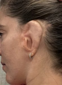
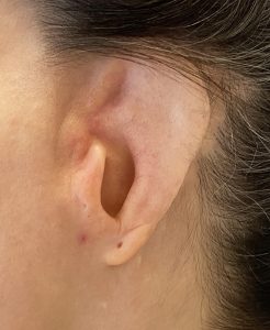
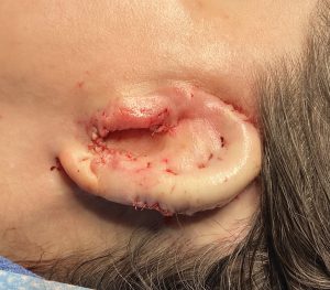
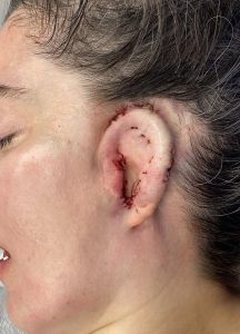 The immediate results of the secondary ear reconstruction showed a better shaped and more proportionate ear size.
It became apparent once the ear framework was uncovered that the outer helical rim graft during the original microtia surgery had slipped off the framework, resulting in an unintentional elongation of the ear. This also explains why there was limited helical rim show of the upper half of the ear framework. This provided the opportunity for making a significant change in the size and shape of the reconstructed ear.
Case Highlights:
1) Ear reconstruction with rib grafts in children has been a mainstay of microtia surgery for decades but the effects of growth on the implanted rib tissue has rarely been reported.
2) Microtia surgeries that end up as macrotia (ears that are too vertically big) can be improved by secondary reconstructive techniques.
3) Most secondary reductive microtia reconstructions can be done without the need for additional tissue graft harvests.
Dr. Barry Eppley
Indianapolis, Indiana