

Background: While the skull is generally a collection of smooth convex surfaces, it is prone to a wide variety of contour deformities. Many of these contour deformities are located in and around the suture lines which serve as the interface between two growing skull bone plates. As there are six primary sutures on the external skull (coronal, sagittal, lambdoid) there is a lot of opportunity for surface contour irregularities to occur.
The coronal suture separates the parietal bone plates from the frontal bone plate and runs across the top of the head from side to side. Where the coronal suture meets the midline sagittal suture is known as the bregma. It is the back edge of the anterior fontanelle which is open at birth and closes around 18 to 24 months after birth. Premature fusion of this suture line creates well known craniosynostosis head shape deformities.
But there are numerous less significant shape issues that can occur around the coronal suture that are not blatantly synostotic. One of these is seen when an indentation occurs along the coronal suture line creating a dip or indentation along its course. It can occur on one side up to the bregma or can occur on both sides across the bregma. This may represent some form of incomplete suture synostosis although this has never be proven.
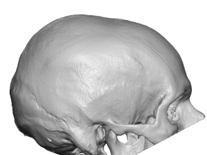
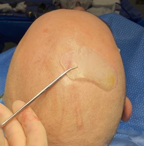
In the shaved head male coronal dip deformities are very apparent and makes the head appear like it has a dent in it.
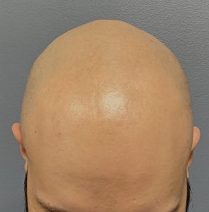
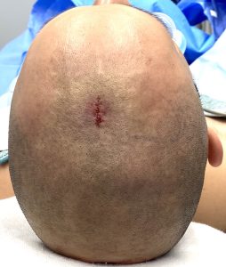
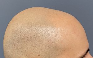 Case Study: This shaved head male presented for the treatment of a noticeable indentation the right side of the top of his head. It occurred along what was believed to be the coronal suture line.
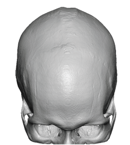
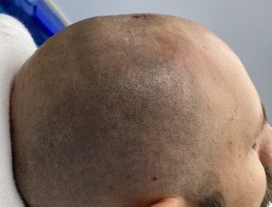
 A 3D CT skull scan confirmed that the indentation was along the coronal suture line with a visible raised bony edge at the front of the indentation near the midline.

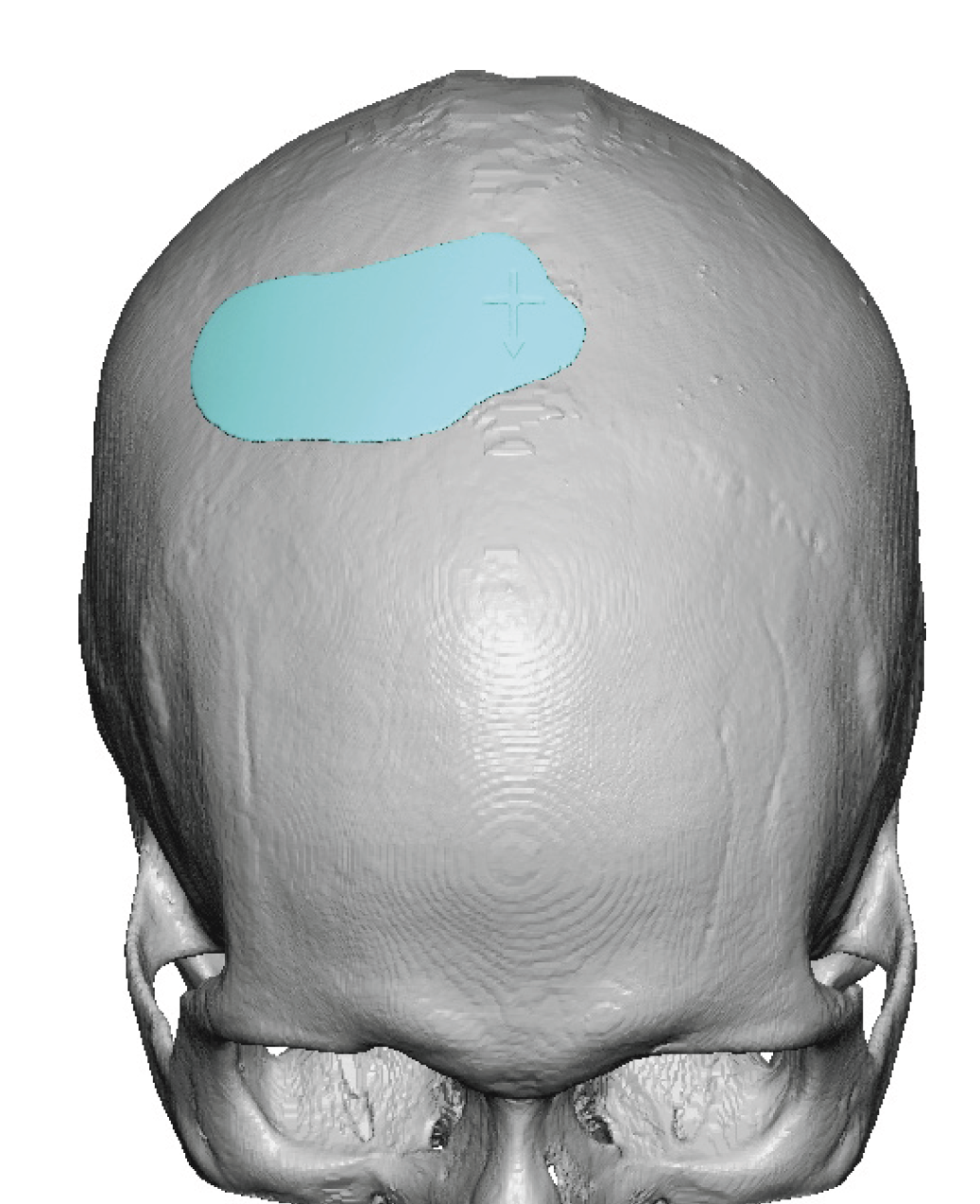
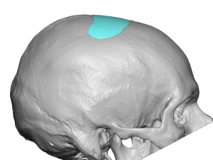 A custom skull implant was designed using a mirroring technique based on the other side. His aesthetic concern was to avoid any risk of overcorrection so it was made smaller than what what the mirroring technique initially showed.
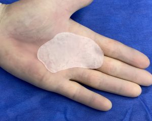
 Its thickness was 2mms at its maximum with a volume of 2ccs.
 Under general anesthesia a 1 cm midline scalp incision was made over the sagittal suture line at the edge of the indentation. Through this incision a subperiosteal pocket was developed into which the implant was placed.
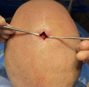
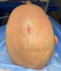 It was positioned and secured with a single 1.5 x 4mm micro screw. A two layer resorbable scalp closure was done.
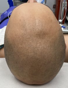
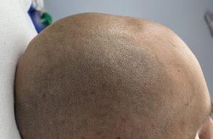
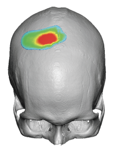
 His intraoperative results shows the elimination of the visible coronal dip indentation on the right side.
The coronal dip is a small indentation that creates a visible dent in the shaved head male. Its presence is magnified by light reflections off of the smooth scalp surface. It is ideally treated by a custom skull implant which avoids the risk of implant augmentation edging as well as over correction.
Case Highlights:
1) Indentations along the coronal suture lines on the top of the head can occur on one or both sides of the sagittal suture line and are known as coronal dip skull deformities.
2) A small custom made implant is the most accurate method to treat the small coronal dip skull indentations.
3) Placement of small skull implants can be done through remarkably small incisions placed right next to it.
Dr. Barry Eppley
World-Renowned Plastic Surgeon