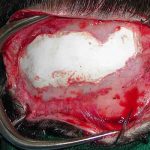
EXPLORE
Plastic Surgery
Dr. Barry Eppley

Explore the worlds of cosmetic
and plastic surgery with Indianapolis
Double Board-Certified Plastic
Surgeon Dr. Barry Eppley

Archive for the ‘cranioplasty’ Category

Cosmetic Skull Contouring With Occipital Augmentative Cranioplasty

Tuesday, July 26th, 2011

When one thinks of cosmetic surgery, the thought rarely goes to any form of skull contouring. While the world is full of a wide variety of head shapes, there is no uniform size or measurement to determine a pleasing cranial contour. But there is a certain oblong and curvilinear shape to most of the skull. The forehead, which is really the front part of the skull, has some very specific desired shapes and contours that are very gender-specific, but the rest of the skull has less well-defined determinants of being pleasing in appearance.

Despite the comparatively obscure location of the back of the head (occiput) compared to the forehead, not everyone is entirely pleased with this portion of their skull shape. The typical concern is that the back of the head is too flat without adequate projection. Whether it be the entire occiput or limited to just the crown area (upper occiput), this is a legitimate cosmetic concern for those so affected. For anatomic clarification, the bony portion of the occiput actually stops at about the level of the middle of the ear. The rest of the back of the head is muscle and other soft tissue.

The most effective method of occipital skull contouring is augmentative, putting a material on top of the bone to build it out. This is a common form of cranioplasty that has a long history in the forehead, it is just less commonly done on the back of the head. There are multiple ways to perform an occipital augmentation cranioplasty and they differ primarily in the material used and the incisional approach to get it there. Each has their own distinct advantages and some disadvantages.

The open form of an occipital cranioplasty uses a straight line vertical incision of about 3 or 4 inches down the back of the head. This approach heals better than any other scalp incision in this area. The scalp is lifted off of the bone around a premarked area of the flattening. Through this approach, either an acrylic (PMMA) or calcium hydroxyapatite (HA) material can be mixed, placed and contoured to the desired shape. Both are powders and liquids that are mixed together to form a moldable putty which as about 10 minutes of working time. Because it is a putty, thus is why it needs an open approach to be placed. Acrylic has the advantage of setting up as solid as bone and just as fracture-resistant. That would offer a theoretical advantage on the back of the head since we lay on it all the time. HA, which setting up firm, is a bit more brittle and fracture-prone although I have never seen that to be a problem in the forehead. With either material, the open approach offers the best chance for a very smooth and even contour shape.

The limited incision or injectable approach uses a small one inch incision through which a material is injected by a small tube. (not a needle) The only cranioplasty material that has the physical properties once mixed (for the first 4 minutes) to flow through a small tube is Kryptonite. (KP) This is a calcium carbonate material that is also highly compatible and will allow some skull bone ingrowth. It does not get as hard as PMMA but it slightly less brittle than HA. The disadvantage to the injected material approach is that there is a higher risk of contour irregularities and palpable edges. This occurs because the molding of the material as it sets is done from the outside so there is no visual way of confirming how smooth it is as it sets.

Cosmetic contouring can be done on most areas of the skull. Occipial augmentation cranioplasty can be done to build out a flat spot or entire back of the head. Regardless of the material and the incision used, it is a simple procedure for a patient to go through with very minimal recovery. Patients report having a headache for a few days but no real pain. Once can wash their hair within 48 hours. Some mild swelling can be expected and the final result can be fully appreciated within 6 weeks after the procedure. All of the cranioplasty materials used are permanent so the change in skull contour will be maintained over one’ lifetime.

Dr. Barry Eppley

http://www.eppleyplasticsurgery.com

Indianapolis, Indiana

Screw Fixation of Acrylic Onlay Cranioplasty

Tuesday, July 19th, 2011

One of the techniques for contouring of the skull is augmentative cranioplasty. This is where the skull or forehead is built out through the use of a variety of synthetic materials. While bone would seem to be a logical choice, it is associated with resorption when used as an onlay not to mention the need for a donor site. Synthetic materials offer contour stability, off-the-shelf ease of use and the elimination of a donor site. Even though there is a risk of infection with synthetics, their benefits far outweigh this very uncommon cranioplasty risk.

One of the most common cranioplasty materials is acrylic or PMMA. (poly methylmethacrylate) It has been used successfully for decades and offers a material strength that is similar to bone and is virtually impact-resistant. It is used in orthopedics as a bone cement for joint prostheses but on the skull it does not have ‘cement’ capabilities. This means that the material does not bond to the bone or the overlying soft tissues. Rather it forms a scar capsule around the material, particularly between the material and the underlying bone. This means that there can be small amounts of motion or instability of the cranioplasty material. In revision of pure onlay cranioplasties, it can be very easy to lift the material off of the bone. This material looseness may or may not cause any long-term problems.

Since a PMMA cranioplasty does not bond to the bone, I have always used an anchoring method for the material. The simplest method is to preplace small titanium crews into the outer table of the skull and leave them raised several millimeters. When the cranioplasty material is applied, the initial putty phase of the material wraps around and grabs the screwheads. Once cured, there is a rigid lock of the material to the bone. This prevents any chance of instability or shifting of the material on the skull’s surface.

This screw anchorage of cranioplasties can be used at any location on the skull. It is of particular value on the occipital (back of the head) region where the skull is exposed to the greatest amount of regular stress. (laying on the back of your head)

This cranioplasty fixation method is not as important with the use of other materials such as hydroxyapatite. While these materials are far weaker and minimally impact-resistant, they do have the ability to bond to bone so they have no risk of material movement or instability.

Dr. Barry Eppley

http://www.eppleyplasticsurgery.com

Indianapolis, Indiana

Case Study: Secondary Hydroxyapatite Cranioplasty Reconstruction

Thursday, April 7th, 2011

Background: The forehead occupies the upper third of the face and has variable degrees of convexity based on gender and genetics. It is structurally composed of very strong bicortical bone at its upper two-thirds and the much weaker aerated brow bones at its lower third underneath the eyebrows. While the forehead can resist significant forces, up to more than 100kgs of impact, it can be fractured and displaced. The brow bones, however, fracture at much lower impact forces.

Because it takes such a tremendous force to fracture the frontal bone, most such injuries also cause brain trauma and frequently need neurosurgical intervention. Often a craniotomy is done and the forehead bone is reconstructed at that time. But some cases do not involve the need for neurosurgery and the forehead defect is lefted untreated. These forehead contour defects are then reconstructed secondarily.

Case Study: This is a 37 year-old male who sustained significant forehead trauma and fractures from a fall from a scaffolding at his work one year previously. He recovered from his head trauma and had full neurological recovery. He had a large indent in his forehead and a well-healed vertical laceration that extended from his hairline down to his eyebrow. A CT scan shows the degree of outer cortical table impaction from his frontal sinus upward.

An open cranioplasty was performed by re-opening his healed laceration. Through this approach the forehead tissues could be lifted back onto the normal undepressed contours of the bone in a circumferential manner. The infractured bone was largely healed and there was no communication into the frontal sinus cavity. An hydroxyapatite paste was prepared, packed into the defect and made smooth with the surrounding bone. In less than 10 minutes, the hydroxyapatite paste hardened and the incision closed.

He had complete restoration of his forehead contour and its convexity and his incision healed without problems. This procedure was done as an outpatient and completed in less than 90 minutes. His postoperative pain and forehead swelling was minimal.

The hydroxyapatite cranioplasty concept is now about 15 years old in its clinical use. It offers an advantage over old-style PMMA (acrylic) in that the material is more biocompatible and actually integrates into the tissues. It is more expensive than PMMA and is far less impact/fracture resistant.But I have never yet seen that be a secondary cranioplasty problem. Due to the handling of the material it must be placed through an open approach. In some cases, small incisional access can be used to place the material.

Case Highlights:

1) Forehead defects from trauma involve changes in the contour of either the solid portion of the forehead and/or the more fragile brow bones.

2) Contour reconstruction of the depressed forehead can be done with the onlay cranioplasty material, hydroxyapatite, in an open technique.

3) Frontal cranioplasty can be done through either a coronal (scalp) incision or an existing forehead laceration if it is big enough.

Dr. Barry Eppley

http://www.eppleyplasticsurgery.com

Indianapolis, Indiana

Case Study: Minimally Invasive Cranioplasty using Kryptonite Bone Cement

Wednesday, October 13th, 2010

Background: The shape of one’s skull is not often thought of as a major cosmetic issue. Much of this is because it is largely hidden by hair in most women and in many young men. But with hair loss in men and the contemporary hairstyle of total scalp shaving, the shape of one’s skull and upper forehead can take down a more dominant visible role. The overall size of the skull is actually greater in surface area than that of one’s face. And, unlike the face, it is generally desirous to be more of a smooth oval rather than have a lot of indentations and irregularities.

Reshaping of one’s skull can be done by either bone reduction or augmentation by onlay materials. By far, more significant changes can be made in the shape of an adult’s skull through addition rather than subtraction. Such cranioplasty procedures have been historically done through an open scalp (coronal) approach. By making a long transverse scalp incision, the scalp tissues can be widely elevated providing wide open exposure and an unobstructed working field. When one has a skull deformity as a sequelae of a prior neurosurgery or craniofacial procedure, the use of a pre-existing scalp incision makes the decision easy to proceed with skull contouring. In the absence of a prior scalp scar, however, the decision to undergo any form of skull reshaping is complicated by the scar’s cosmetic trade-off.

Case Study: A 25 year-old male wanted skull and forehead reshaping due to some cosmetic irregularities. He had a pair of grooves or indentations on each side of the midline of his skull that ran from the back of his head to the middle of the forehead. These depressions were visible given his receding hairline and close cropped hair. These parasagittal linear indentations were somewhat reminiscent of a ‘miniature’ or micro- form of sagittal craniosynostosis as noted by a midline ridge along the sagittal suture and restricted bone growth perpendicular to it along its length. He has trouble wearing hats as his narrow forehead allowed the hat to fall down unsupported to his brows.

These parasagittal skull grooves could be easily augmented through a traditional open cranioplasty approach. Understandably he did not want the scalp scar given the modest nature of the skull problem. An alternative approach of an injectable cranioplasty procedure was devised that could be done through very small incision but yet could add material over the entire lemgth of the skull defects. Such an approach, while not possible with the use of traditional cranioplasty materials such as acrylic and hydrozyapatite, can be done using the newly FDA-approved Kryptonite bone cement.

Prior to the procedure, an outline of the skull depressions were marked on both sides. Clay modeling material was used to fill in the defects and shaped. They were used to give an estimate of how much material would be needed to fill in the defects from front to back. The patient was taken to the operating room where a small one inch incision was made at the back end of the linear  depressions. Long curved elevators were used to raise the scalp tissue off of the bone in a subperiosteal plane within the confines of the skin marks. The pocket was the washed out with antibiotic solution. On each side, 10 grams of Kryptonite bone cement were then injected from front to back using a syringe and a long 12 French plastic catheter. The material was shaped externally by molding with fingers to get smooth edges and sn even fill of the skull depressions to the surrounding skull bone. Once set after 10 minutes, the incisions were closed with small dissolveable sutures. Total operative time was one hour and was done as an outpatient procedure

When seen the next day, he reported only a mild headache for a few hours after surgery. He took no pain medications at all. His swelling was mild as expected and he had no bruising. The effects of the skull contouring was immediately evident and pleasing. However, swelling remains and the final outcome of the procedure will take several months before its true success can be judged. What the procedure does illustrate is that it is possible to do select cranial augmentation using this method. It is fairly simple and the patient will have a quick recovery.

For more modest skull contour deformities where a long scalp incision would be cosmetically unacceptable, the injectable cranioplasty method is a viable method. It is made possible with the flow characteristics of Kryptonite bone cement that have not previously existed with prior cranioplasty materials.

Highlights:

1)      Skull irregularities and contour depressions have traditionally used an open scalp incision with its own cosmetic scar trade-off.

2)      Injectable cranioplasty uses very small incisions that can fill in a large surface area through a long flexible filler tube. It is a procedure that is technique sensitive and intimate working knowledge of how the Kryptonite material flows and sets, and the time frame on which it happens, is needed to ensure a good result.

3)      Cosmetic skull reshaping by augmentation is now possible with the minimally invasive cranioplasty (MIC) procedure. Further work is needed to define the procedure to avoid undercorrection and, most significantly, any risk of overcorrection.


Dr. Barry Eppley

 http://www.eppleyplasticsurgery.com

Indianapolis, Indiana

Kryptonite Bone Cement - A Potential Injectable Cranioplasty Method

Monday, July 26th, 2010

Cranioplasty is done for making contouring changes to the cranial vault, which is defined as the skull, forehead and brow bones. Common causes for the need for cranioplasty are congenital deformities (after primary reconstruction), neurosurgical bone flaps, and traumatic injuries. When done for these reasons an open approach is always used as there is usually a pre-existing scalp scar/incision from a prior procedure. This makes it very easy to apply the traditional synthetic cranioplasty materials such as PMMA (acrylic) and HA. (hydroxyapatite)


However, some skull shape problems may be relatively small or may not be associated with any pre-existing incision for access. When balancing the trade-off of a new scalp scar versus keeping the existing skull concern, many patients (particularly men) would consider the scar as more undesireable. Cranioplasty would be more appealing in this circumstance if the cranioplasty material would  be able to be delivered from small and remote incisions. In essence, a cranioplasty material that could be delivered by an injection process.


The current craniplasty materials are far from ideal to be delivered through any form of remote access. PMMA, polymethylmethacrylate, is an initial liquid which can be delivered through a tube but it is very runny on delivery and sets up with a very high heat from an exothermic reaction. The numerous forms of hydroxyapatite (e.g., Mimx) create an initial viscous slurry which has no material flow at all. This makes it not only undeliverable by injection but its sensitivity to fluids and its easy fragmentation on setting make external digital molding unpredictable.


Kryptonite, the newest FDA-approved cranioplasty material offers some real promise as an injectable skull-shaping technique. It is a bone cement that is created through the combination of a calcium carbonate powder and two fatty acid liquid derivatives from castor oil. When combined together this makes a final hardened material that is porous and adhesive with bone-like properties. While this is as favorable to bone as any of the other cranioplasty materials, its physical features in the set-up process make it potentially useful to be delivered by an injection method.


Once the three ingredients are mixed, a very flowable liquid is created. During the intial polymerization process (up to 4 minutes), the material can be loaded into a syringe. Once in the syringe, it remains in a thick but flowable liquid phase up to 8 minutes are mixing. This provides the opportunity for delivery by injection. Once it passes the 8 minute time period, it enters a sticky taffy phase where it becomes very adhesive and is no longer injectable. A moldable phase will exist then up to 25 minutes in which further shaping can be done.


To pass the injectable cranioplasty test, it first must have enough flow to be delivered through a small enough tube that has sufficient length. A true injection method  is delivered percutaneously through a needle that is no larger than an 18 gauge. But that is not the type of injection to which I refer when being used for cranioplasty. Because instruments must be used to develop the subperiosteal pocket, either through an endoscopic or blind technique, one or two small remote incisions (less than 1 inch) would be used. Therefore the internal diameter of the injection method can be larger. But how large does it need to be? (or how small can be that it still works) In bench top testing, I have determined that it flows very nicely through an internal diameter of 2.7mms. (8 French catheter) This makes it possible to use long catheters for remote access. Some material will be lost in the tube during delivery which is 1cc per 13cm of length at 2.7mms diameter. Most injectable cranioplasties will not need more than 5 or 6cms of tube length for delivery. The indwelling .5cc of material can be pushed through with a saline fluid bolus behind it.


The other injectable consideration is can the material be effectively molded by external manipulation. Is it able to be pushed around and molded into fine edges without fracture or separation of the material? In testing on a pig’s head (from the butcher), Kryptonite was injected and externally molded to the back of the skull from an anterior incision. On dissection after setting, it was adherent to the bone, did not stick to the overlying soft tissue and had nicely contoured edges. Its sticky taffy phase which is between 5 and 15 minutes after mixing gives it a texture which really molds and shapes well.


Kryptonite bone cement appears to offer physical properties that would make it the first truly injectable cranioplasty material. Its use in this manner is for partial-thickness contour deformities of the skull and forehead.       


Dr. Barry Eppley

http://www.eppleyplasticsurgery.com

Indianapolis, Indiana  


Dr. Barry EppleyDr. Barry Eppley

Dr. Barry Eppley is an extensively trained plastic and cosmetic surgeon with more than 20 years of surgical experience. He is both a licensed physician and dentist as well as double board-certified in both Plastic and Reconstructive Surgery and Oral and Maxillofacial Surgery. This training allows him to perform the most complex surgical procedures from cosmetic changes to the face and body to craniofacial surgery. Dr. Eppley has made extensive contributions to plastic surgery starting with the development of several advanced surgical techniques. He is a revered author, lecturer and educator in the field of plastic and cosmetic surgery.

Read More


Free Plastic Surgery Consultation

*required fields


Pricing

The cost of any type of elective plastic surgery plays a major role in the decision to undergo the procedure(s).

More Info


Military Discount

We offer discounts on plastic surgery to our United States Armed Forces.

More Info


Categories