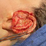
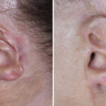
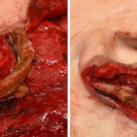
EXPLORE
Plastic Surgery
Dr. Barry Eppley

Explore the worlds of cosmetic
and plastic surgery with Indianapolis
Double Board-Certified Plastic
Surgeon Dr. Barry Eppley

Archive for the ‘ear reconstruction’ Category

Case Study: Rib Graft Reconstruction of the Avulsed Ear

Thursday, February 3rd, 2011

Background:  One of the most challenging of all plastic surgery procedures of the face is ear reconstruction. Microtia, or congenital absence of the external, is the most extreme challenge when it comes to making an ear essentially from scratch. A more common, although not less challenging, is that of partial ear reconstruction.

Whether it be from an automobile accident, a sharp edged instrument or even a bite wound, the loosely attached and flexible cartilage of the ear and its attached skin is relatively easy to avulse or be amputated. Loss of part or all of the ear cartilage, while not life-threatening, is nonetheless disfiguring and very psychologically disturbing.

I have seen ear amputations present numerous times with the avulsed ear segment in hand (or someone else’s hand) with the understandable hope that it may be able to be reattached. This is rarely possible although there is no harm in making an attempt if it is not overly crushed or mutilated, which it often is. Unless the ear is in fairly pristeen condition, it is better to close the wound and let it heal for six months before embarking on ear reconstruction efforts.

Case Study: This 18 year-old female presented in the emergency room from a rollover automobile accident on the night of her high school prom. The avulsed ear segment was brought in by the ambulance crew but it was completely crushed and unusable. She was taken to the operating room where her ear wounds were closed.

Six months later she presented for ear reconstruction. Initially tissue expansion was planned as the first stage prior to rib graft placement but she needed to complete her ear reconstruction as possible. (she was enrolling the military)

In her definitive one-stage ear reconstruction, the first step is to make a template of the opposite ear and transpose it to the amputated ear site to get the right shape and orientation of the reconstruction.

Rib grafts were then harvested using the confluence of rib numbers 6 and 7 as well as number 8 to make the helix. These were then carved and put together to make a framework for the ear reconstruction. This creates a complex framework of concavities and convexities which mimic those of the natural ear.

Because of her tight scar and skin coverage over where a rib graft reconstruction was to be placed, a temporoparietal fascial vascularized flap was planned. This was raised to be large enough to turn down and cover the full extent of the rib graft.

The rib graft is then placed and the temporoparietal flap used to provide vascularized coverage over it. A split-thickness skin graft, harvested from a non-hair bearing area, is then placed over it and meticulously sutured down. A compressive dressing is applied afterwards over drains placed underneath the cartilage framework.

The skin graft went on to have a 100% take over the combined rib graft and fascial flap. There remained a slight pigmentation difference in the skin graft from that of her natural ear and neck skin.

Case Highlights:

1)      External ear reconstruction from partial or total amputation injuries is best done with rib cartilage when significant portions of the framework are missing.


2)      Due to scarring and skin loss, more skin has to be created to produce an ample and supple skin cover over the rib graft. This is most commonly and easily done with tissue expanders which makes it a staged reconstruction approach.


3)       A one-stage ear reconstruction approach can be done using a temporoparietal fascial flap and split-thickness skin graft coverage.


Dr. Barry Eppley

http://www.eppleyplasticsurgery.com

Indianapolis, Indiana

Microtia Ear Reconstruction - A Plastic Surgery Marvel

Wednesday, July 22nd, 2009

Microtia is a well known congenital ear deformity that has a broad spectrum of presentations. From a small residual bump to a crumpled shortened ear, no two cases of microtia are exactly alike. But they all similar in that they are an incompletely formed ear that is much smaller than a normal one. The underlying cartilage framework and ear canal are malformed and it is easy to see why such a complex structure can go developmentally awry.


One of the marvels of modern plastic surgery is the reconstruction of the microtia ear deformity. While it is not a life-threatening or life-changing deformity, its reconstruction is a complex array of operative steps that call upon a variety of basic plastic surgery techniques and an artistic ability. While it is not as technically precise as the vascular anastomosis of a free flap, it is similarly unforgiving in its execution. You have one good opportunity to work with the skin over the deformed cartilage remnant. Your first effort is your best chance for the best result. Salvages of failed ear reconstructions are extremely difficult and almost always far from satisfying.


While there are different methods for putting the initial cartilage framework together, and it is a learned skill to masterfully assemble one, the more critical step is in how the overlying skin is handled. What is the correct location and angulation of the framework, is the skin adequate or does it need tissue expanded,  how much of the deformed cartilage should be removed, and what about the typical low lying hairline? All of these issues can compromise an otherwise well-crafted cartilage framework by obscuring its exquisitely carved detail.


Beyond the most critical step of first-stage cartilage framework fabrication and insertion is the creation of smaller but aesthetically important ear details. The lobule, ear to head separation, and the concha-tragus unit all make important contributions to what we see as a more normal ear. While different microtia surgeons may stage these subsequent creations at different times or in differing combinations, all will attest to their value in the pursuit of what nature failed to create.


Even with all the current and future innovations of plastic surgery, microtia reconstruction remains a marvel and is a collage of plastic surgery techniques focused one relatively small area. The sheer rarity of microtia makes its reconstructive mastery limited to just a handful of plastic surgeons.


Dr. Barry Eppley

http://www.eppleyplasticsurgery.com
http://www.ologyspa.com
Clarian North Medical Center, Carmel, Indiana
Clarian West Medical Center, Avon, Indiana
Indianapolis


The Architecture of Ear Plastic Surgery

Wednesday, April 2nd, 2008

The ear may be quite small, measuring only about 5 cms in height and 3 cms in width, but it has the most complex anatomy of any facial component. Its many ridges and convolutions comprised only of cartilage (and the overlying skin), surrounding the ear hole, give it a distinct shape that is uniquely recognizeable as an ear. But within its complex geometries lie some basic architecture that guides how otoplasty (ear pinning) or ear reconstructive surgery is done.
On a simplistic level, the ear is three levels or tiers. An amphitheatre that encircles a central stage if you will. The outer or top layer is the outer rim of the ear known as the helix and it sits the highest. (farthest away from the side of the head) The next step down is an inner rim or antihelix which parallels the helix for the most part but at a lower level. And the final step down into the hole, so to speak, is the bowl or concha. Understanding the three tiers or levels of the ear is to understand how to surgically change it.
In otoplasty, often called ‘pinning back the ears’, the anithelix is missing. (the fold is not there) So to move the ears back, the cartilage from behind is sewn closer together to make an antihelix or antihelical fold, thus moving the helix and ear back closer to the head. How snug or loose these shaping sutures are placed determines how close the ear sits to the side of the head. In some cases of protruding ears, the bowl or concha is also too big. So the concha from behind may be cut down in size by cutting out a wedge or sewn directly back, this also moving the ear back. Since otoplasty is mainly about shaping the cartilage with sutures, this is why it is a simple and fairly quick operation…but with a very powerful visual effect.
Conversely, ear reconstruction can be quite complex. In children born with much or all of their ear missing (known as microtia), complex cartilage grafting must be done. This often involves taking rib cartilages, putting them together, and carving out an ear framework. And how is the framework pieced together and carved? Based on the three-tier principle of ear architecture! Helix, antihelix, and concha.While ear reconstruction is multiple stages and is not based solely on the cartilage framework that is put under the skin, it all begins with a well-fabricated cartilage framework. Onto that are finer details of the lobule and other shaping procedures (more minor) which are done later.
Dr Barry Eppley
http://www.eppleyplasticsurgery.com/
http://www.ologyspa.com/
Clarian North Medical Center, Carmel, Indiana
Clarian West Medical Center, Avon, Indiana]
Indianapolis

Surgical Correction of Ear Gauging

Saturday, February 23rd, 2008

One of the recent trends of personal marking or adornment is that of gauging. Similar to piercings, gauging is basically an extension of this concept only with an end result of larger holes and jewlery. It is done by starting with a traditional piercing, usually in the ear, and then gradually enlarging the piercing hole by the slow sequential replacement of a ‘hole expander.’ Over time, the earlobe hole gets stretched until it can accomodate a metal insert that is often larger than the original size of the earlobe.
Gauging is undoubtably a short-lived phenomenon done mainly in the young. Eventually, most people with gauging piercings or jewelry may want it reversed. Fortunately, in the ear, this is fairly easy to do. Because no actual earlobe skin has been removed, but merely stretched, its correction is similar to that of an earlobe reduction procedure. The edges of the enlarged hole are excised and brought together, restoring the earlobe to a near normal size.
In areas other than the earlobe, however, gauging is not so easily corrected. In other parts of the ear, there would be loss of cartilage (unlike the earlobe which is only made made up of skin) and this can be replaced. Such holes in cartilage-containing part of the ear must be cut out with significant alteration of the size and shape of the ear to close it.
Dr Barry Eppley
http://www.eppleyplasticsurgery.com
http://www.ologyspa.com
Clarian North Medical Center, Carmel, Indiana
Clarian West Medical Center, Avon, Indiana
Indianapolis

Otoplasty (Ear Setback) Surgery

Sunday, November 11th, 2007


While there are many other plastic surgery procedures that are more popular, one of the most simple and dramatic procedures is that of otoplasty. Performed in around 20,000 Americans last year, it has one of the most dramatic effects on self-image of any operation in plastic surgery. Usually done in younger patients (most commonly under age 18), often those teased throughout school due to their ‘dumbo’ ears, the ears are reshaped so that they lay closer to the side of the head.


The operation is done by cutting on the back of the ears, exposing the back surface of ear cartilage. Special sutures are placed to bend the right area of the cartilage to create a helical rim (outer aspect of the ear) that sits back further. The tightening of the sutures determines how far back the ear is set. You don’t want to overtighten these sutures. If you do, you will create the classic ‘telephone-ear’ deformity. (the ear plastered against the side of the head) Overcorrection is impossible to fix later, undercorrections (while also not desireable) can at least be set back further later.


In children, the procedure is done under general anesthesia in the operating room. In select adults, it can be done under local anesthesia in the office. While in young children (under age 8), I put a head dressing on for a week, in adults I only use it for one day. While some ear swelling is seen for a few weeks, the surgical results are immediately apparent. One other benefit, there is some ear soreness but no real acute pain with the procedure.


I often joke that many otoplasties are done in children in the spring, when the winds pick up.

Dr Barry Eppley
www.eppleyplasticsurgery.com
www.ologyspa.com
Clarian North Medical Center, Carmel, Indiana
Clarian West Medical Center, Avon, Indiana
Indianapolis


Dr. Barry EppleyDr. Barry Eppley

Dr. Barry Eppley is an extensively trained plastic and cosmetic surgeon with more than 20 years of surgical experience. He is both a licensed physician and dentist as well as double board-certified in both Plastic and Reconstructive Surgery and Oral and Maxillofacial Surgery. This training allows him to perform the most complex surgical procedures from cosmetic changes to the face and body to craniofacial surgery. Dr. Eppley has made extensive contributions to plastic surgery starting with the development of several advanced surgical techniques. He is a revered author, lecturer and educator in the field of plastic and cosmetic surgery.

Read More


Free Plastic Surgery Consultation

*required fields


Pricing

The cost of any type of elective plastic surgery plays a major role in the decision to undergo the procedure(s).

More Info


Military Discount

We offer discounts on plastic surgery to our United States Armed Forces.

More Info


Categories